





The integrity of the skeletal system relies on a dynamic balance between formation and resorption processes. Within this balance, even microscopic alterations can influence overall bone strength. Understanding how microdamage accumulates is essential for clinicians and researchers seeking to prevent fractures and optimize therapeutic interventions.
Bone Microdamage: Fundamental Concepts
Bone tissue endures repetitive loads and physiological stresses throughout life. Tiny cracks, known as microcracks, develop when mechanical demands exceed local material capacity. These microcracks coalesce or propagate under cyclic loading, potentially leading to macrostructural failure. Researchers classify microdamage into two major categories: linear microcracks and diffuse damage. Linear microcracks appear as discrete fissures in the collagen-mineral matrix, whereas diffuse damage comprises clusters of submicroscopic cracks that reduce local stiffness without a visible fracture line.
Bone microdamage primarily originates from everyday activities such as walking, running, or lifting. The process closely parallels mechanical fatigue in engineering materials, where repeated stress cycles trigger progressive deterioration. Unlike inert alloys, bone possesses a unique ability to sense and repair damage via cellular pathways.
Cellular Mechanisms and Remodeling Response
At the heart of microdamage detection lie osteocytes, the most abundant bone cells embedded within mineralized matrix compartments called lacunae. Osteocytes form an interconnected network through canaliculi, allowing them to monitor mechanical strains and fluid flow. Upon identifying excessive deformation or fluid shear stress, osteocytes release signaling molecules—such as sclerostin, RANKL, and nitric oxide—to recruit remodeling units.
Bone remodeling unfolds in a coordinated sequence:
- Activation of osteoclast precursors and localized resorption.
- Formation of Howship’s lacunae where old tissue is replaced.
- Recruitment of osteoblasts to deposit new collagen and initiate mineralization.
This cyclical process maintains skeletal integrity by removing microdamaged regions and replacing them with fresh, structurally sound bone. However, aging, hormonal imbalances, and metabolic disorders may impair this reparative capacity, allowing damage to accumulate over time.
Factors Influencing Microdamage Accumulation
Several intrinsic and extrinsic factors modulate the rate at which microdamage accrues:
- Bone Density: Lower mineral density reduces the threshold for crack initiation.
- Material Properties: Alterations in collagen cross-linking or mineral crystal size affect toughness and brittleness.
- Loading Patterns: High-impact or repetitive activities increase the frequency of damage events.
- Vascularization: Adequate blood supply ensures efficient removal of necrotic debris and supports osteocyte viability.
- Nutrition and Hormones: Deficiencies in calcium, vitamin D, or estrogen accelerate bone turnover and remodeling imbalances.
Biomechanical studies simulate various loading scenarios on animal or human specimens. Finite element analysis models highlight stress concentrators—such as cortical pores or osteonal boundaries—that predispose specific regions to microcrack formation. Understanding these patterns aids in targeted prevention strategies for at-risk populations.
Diagnostic Techniques and Imaging
Detecting microdamage in vivo poses significant challenges due to the scale of defects. Advances in imaging and histological methods provide valuable insights:
- Fluorescent staining with basic fuchsin reveals microcracks in bone biopsies under microscopy.
- Micro-computed tomography (micro-CT) captures high-resolution 3D images, enabling quantification of damage distribution.
- Nanoindentation techniques assess local biomechanical properties, identifying regions of reduced stiffness corresponding to diffuse damage.
- MRI modalities exploiting ultrashort echo times (UTE) can visualize bound water and matrix alterations adjacent to damage sites.
Integration of these modalities into clinical practice remains limited by cost and accessibility. Yet, they hold promise for early detection of subclinical microdamage before overt stress fractures develop.
Preventive Strategies and Therapeutic Approaches
Mitigating microdamage accumulation requires a multifaceted approach. Lifestyle modifications—such as progressive weight-bearing exercise—enhance bone strength and stimulate adaptive remodeling. Nutritional support ensures building blocks for matrix synthesis, emphasizing adequate protein, calcium, and vitamin D intake.
Pharmacological interventions target remodeling pathways:
- Bisphosphonates: Inhibit osteoclast-mediated resorption, reducing excessive turnover but potentially suppressing normal repair if overused.
- Denosumab: Monoclonal antibody against RANKL, decreasing osteoclast activity with reversible effects on bone turnover.
- Parathyroid hormone (PTH) analogs: Stimulate osteoblast function, promoting new bone formation and microdamage repair.
Emerging therapies focus on modulating osteocyte signaling and improving local microenvironmental conditions. For instance, sclerostin inhibitors enhance Wnt signaling, boosting osteoblast activity and fortifying microstructure. Gene therapy approaches targeting extracellular matrix proteins may one day reinforce bone’s innate resistance to crack initiation.
Future Directions in Bone Microdamage Research
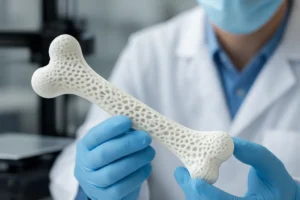
Combining multi-scale computational models with experimental data will refine predictions of microdamage progression under varied loading regimes. Material scientists are exploring biomimetic scaffolds that mimic natural bone architecture, potentially guiding in situ regeneration in critical defect sites. Furthermore, personalized medicine approaches integrating genetic, biochemical, and imaging biomarkers aim to tailor prevention and treatment strategies to individual risk profiles.
Ultimately, a deeper understanding of microdamage dynamics promises to revolutionize bone health management, reducing fracture incidence and improving quality of life across the lifespan.