




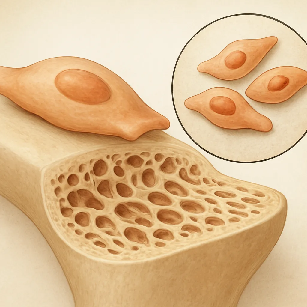
Osteoblasts play a central role in the maintenance, repair, and growth of the skeleton. These specialized cells are responsible for the formation of new bone tissue by secreting and mineralizing the organic bone matrix. Understanding the mechanisms that regulate osteoblast function is essential for developing therapies for conditions such as osteoporosis, fracture nonunion, and metabolic bone diseases.
Physiology of Osteoblast Differentiation
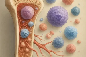
Osteoblasts originate from mesenchymal stem cells (MSCs) within the bone marrow stroma. The process of lineage commitment and maturation involves a series of tightly regulated events governed by transcription factors, signaling molecules, and the local microenvironment.
Origin and Lineage Commitment
Mesenchymal progenitors differentiate into pre-osteoblasts under the influence of key transcriptional regulators. The master regulator RUNX2 is indispensable for the initial commitment phase. Without sufficient RUNX2 expression, MSCs cannot embark on the osteogenic pathway. Later, osterix (also known as SP7) ensures further maturation into fully functional osteoblasts endowed with matrix-producing capacity.
Regulatory Signaling Pathways
A complex interplay of systemic hormones and local factors orchestrates osteoblast differentiation:
- Wnt/β-catenin pathway: Promotes proliferation of pre-osteoblasts and their transition into mature osteoblasts.
- BMPs (Bone Morphogenetic Proteins): Stimulate osteogenic gene expression via SMAD proteins.
- Notch signaling: Modulates the balance between proliferation and differentiation; excessive activation can impair bone formation.
- Hedgehog pathway: Influences early stages of lineage commitment through Indian hedgehog (Ihh).
These pathways converge to regulate the expression of osteocalcin, alkaline phosphatase, and other markers of osteoblast activity.
Matrix Production and Mineralization
Once differentiated, osteoblasts orchestrate the deposition of an organic scaffold and its subsequent mineralization to form rigid bone tissue capable of bearing mechanical loads.
Collagen Synthesis and Secretion
The primary component of the organic matrix is type I collagen, which confers tensile strength. Osteoblasts synthesize collagen molecules in the rough endoplasmic reticulum before secreting them into the extracellular space. Post-translational modifications, such as hydroxylation of proline and lysine residues, are critical for proper triple-helix formation. The resulting collagen fibrils assemble into a highly organized network.
Mineral Nucleation and Growth
Following collagen deposition, osteoblasts create a microenvironment favorable for hydroxyapatite crystallization:
- Matrix vesicles bud from the osteoblast membrane, concentrating calcium and phosphate ions.
- Alkaline phosphatase cleaves pyrophosphate inhibitors, promoting mineral deposition.
- Local pH and ionic strength are regulated to facilitate crystal growth along collagen fibrils.
As mineralization proceeds, the bone matrix hardens, providing structural integrity.
Interaction with Other Bone Cells
Bone is a dynamic tissue maintained through the coordinated actions of osteoblasts, osteoclasts, and osteocytes. Intercellular communication ensures balanced remodeling and adapts bone architecture to mechanical demands.
Coupling with Osteoclasts
Osteoblasts regulate osteoclastogenesis—the process by which bone-resorbing osteoclasts form—via the RANK/RANKL/OPG axis. Pre-osteoblasts and mature osteoblasts express RANKL (Receptor Activator of NF-κB Ligand), which binds RANK on osteoclast precursors, promoting their differentiation. At the same time, osteoblasts secrete osteoprotegerin (OPG), a decoy receptor that sequesters RANKL and inhibits excessive bone resorption. This balance determines the net bone mass.
Communication with Osteocytes
Osteocytes, former osteoblasts embedded within the mineralized matrix, act as mechanosensors. Through a network of canaliculi, osteocytes relay mechanical signals to surface osteoblasts. Mechanical loading suppresses sclerostin, a glycoprotein produced by osteocytes that normally inhibits the Wnt pathway. Reduced sclerostin levels enhance osteoblast activity and accelerate bone formation in response to stress.
Clinical Implications and Therapeutic Strategies
Disruptions in osteoblast function contribute to a variety of skeletal disorders. Targeting osteoblast pathways offers promising avenues for enhancing bone density and accelerating fracture healing.
Osteoporosis Treatments
Current anabolic therapies aim to stimulate osteoblast-mediated bone formation:
- Teriparatide (PTH analog): Intermittent administration increases osteoblast number and activity.
- Abaloparatide: A synthetic analog of PTHrP with similar anabolic effects.
- Anti-sclerostin antibodies (e.g., romosozumab): Inhibit sclerostin to unleash Wnt signaling in osteoblasts.
Enhancing Fracture Repair
In delayed union or nonunion fractures, osteoblast recruitment and function may be insufficient. Strategies include:
- Local application of BMP-2 on collagen carriers to promote differentiation.
- Use of mesenchymal stem cell grafts engineered to overexpress osteogenic factors.
- Biomaterial scaffolds impregnated with growth factors to support extracellular matrix deposition.
Emerging Research in Osteoblast Biology
Recent advances have unveiled novel regulators and potential drug targets within osteoblast signaling networks:
- MicroRNAs: Small noncoding RNAs that modulate osteoblast gene expression post-transcriptionally.
- Epigenetic modifiers: Histone deacetylase inhibitors shown to promote osteoblast differentiation.
- 3D bioprinting: Enables fabrication of complex bone constructs with embedded osteoblast-like cells.
- Gene editing: CRISPR/Cas9 approaches to enhance expression of osteogenic factors or knock out inhibitors.
Understanding how these elements integrate with established pathways will refine therapeutic approaches aimed at optimizing bone health.
Future Directions in Bone Regeneration
The ultimate goal is to achieve controlled, site-specific bone formation without adverse effects. Novel delivery systems for growth factors, tailored scaffolds mimicking the native bone matrix, and patient-specific cell therapies hold promise. Moreover, unraveling the interplay between the immune system and osteoblast function—termed osteoimmunology—may reveal additional layers of regulation critical for both bone development and repair.
Personalized Medicine and Biomarker Discovery
Identifying circulating biomarkers of osteoblast activity, such as propeptides of collagen type I, can inform personalized treatment regimens. Genomic and proteomic profiling will help predict patient responsiveness to anabolic agents and minimize off-target risks.
Integration of Mechanobiology
Advances in wearable technology and in vivo imaging will enable real-time monitoring of mechanical forces and bone adaptation. Coupling this data with osteoblast biology offers the potential to tailor physical therapies that complement pharmacological interventions, maximizing bone strength and resilience.