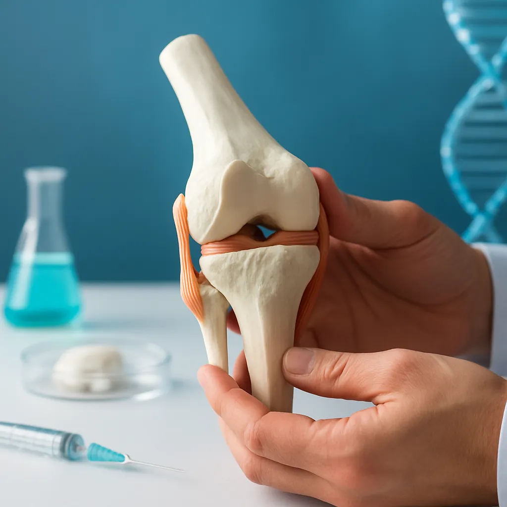



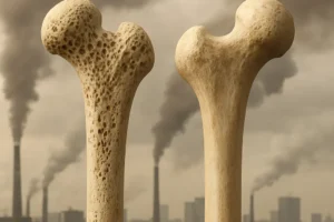
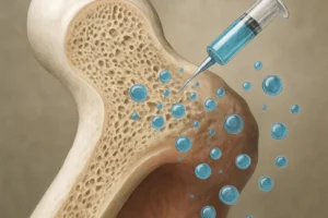
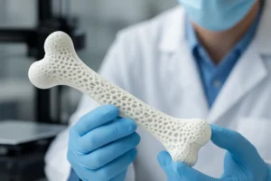
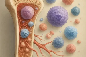
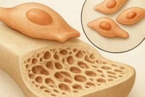


Bone tissue possesses a remarkable capacity for self-repair, yet critical-sized defects and degenerative conditions often exceed its innate healing potential. Recent advances highlight the therapeutic promise of extracellular vesicles (EVs) as a cell-free approach to enhance bone regeneration. These nano-sized particles, secreted by various cell types, carry a rich cargo of proteins, lipids, and nucleic acids that orchestrate intercellular communication during tissue repair. By harnessing EVs derived from mesenchymal stem cells (MSCs) and other sources, researchers aim to stimulate osteogenesis, modulate the immune response, and promote angiogenesis without the safety concerns associated with cell transplantation.
Mechanisms Underlying EV-Mediated Bone Repair
Extracellular vesicles act as natural delivery vehicles, transporting bioactive molecules that influence the behavior of recipient cells. Key mechanisms include:
- miRNA transfer: EVs shuttle specific microRNAs such as miR-21, miR-196a, and miR-218, which regulate critical osteogenic pathways by targeting inhibitors of bone formation.
- Growth factor enrichment: Vesicles are enriched with proteins like BMP-2, TGF-β, and VEGF, fostering both bone formation and new blood vessel ingrowth.
- Immunomodulation: MSC-derived EVs can polarize macrophages toward an anti-inflammatory (M2) phenotype, reducing chronic inflammation and creating a pro-regenerative microenvironment.
- Membrane lipids and surface markers on EVs facilitate receptor-mediated uptake and endosomal escape, ensuring efficient delivery of their cargo.
By engaging cellular signaling cascades—such as Wnt/β-catenin, PI3K/Akt, and MAPK—EVs orchestrate a coordinated response that accelerates matrix deposition and mineralization.
Sources and Isolation Techniques for Therapeutic EVs
A variety of cell types serve as EV donors, each with unique advantages:
- Bone marrow MSCs: High osteogenic potency and well-characterized secretome.
- Adipose-derived stem cells: Abundant yield and ease of harvest.
- Osteoblasts and endothelial cells: Provide lineage-specific signals tailored to bone and vascular regeneration.
Efficient isolation is crucial to obtain high-purity EV preparations. Common methods include:
- Ultracentrifugation: Widely used but time-consuming and may co-isolate protein aggregates.
- Size-exclusion chromatography: Gentle on vesicles and scalable for GMP-grade production.
- Precipitation kits and tangential flow filtration: User-friendly approaches, though purity and yield vary.
Standardizing isolation protocols and characterizing EV preparations—via nanoparticle tracking analysis, electron microscopy, and proteomic profiling—are essential to ensure reproducibility and biocompatibility.
Integration of EVs with Biomaterial Scaffolds
Combining EVs with three-dimensional scaffolds enhances local retention and sustained release, optimizing site-specific regeneration. Strategies include:
- Hydrogel encapsulation: Injectable hydrogels such as gelatin methacrylate (GelMA) or alginate can host EVs, providing a moist environment and controlled degradation.
- Electrospun nanofibers: Polycaprolactone (PCL) or collagen-based mats allow surface adsorption of EVs, mimicking the extracellular matrix architecture.
- 3D-printed ceramics: Composite scaffolds of hydroxyapatite and tricalcium phosphate are functionalized with EVs to promote mineral nucleation and osteoblast adhesion.
These hybrid constructs facilitate sustained release of EV cargo, direct cell migration, and support neotissue formation within critical-sized bone defects.
Preclinical Models Demonstrating Efficacy
Animal studies have provided compelling evidence of EV-induced bone healing:
- Rodent calvarial defect models: Local injection or scaffold delivery of MSC-EVs resulted in significant defect closure and new bone volume, as assessed by micro-CT.
- Femoral fracture models in rats and rabbits: Systemic administration of EVs accelerated callus formation and restored mechanical strength.
- Osteoporotic bone regeneration: EVs derived from young donors reversed age-related impairments in bone healing by enhancing osteoprogenitor activity.
Histological analyses confirm increased collagen deposition, mature osteocyte networks, and enhanced vascularization within treated sites, underscoring the therapeutic potential of EVs.
Engineering EVs for Enhanced Osteogenic Potential
To further boost efficacy, researchers employ bioengineering approaches:
- Genetic modification of parent cells: Overexpression of osteoinductive factors (e.g., Runx2 or BMP-2) enriches EV cargo with pro-osteogenic signals.
- Surface functionalization: Conjugation with targeting ligands—such as RGD peptides or aptamers—improves EV homing to bone injury sites.
- Preconditioning strategies: Hypoxia or mechanical stimulation of MSCs alters EV composition, promoting angiogenesis and matrix synthesis.
- Drug loading: Small molecules (e.g., statins) or nucleic acids are co-encapsulated within EVs to provide multifaceted therapeutic effects.
These advanced methodologies aim to create an “off-the-shelf” product with predictable performance and tailored regenerative capacity.
Regulatory Challenges and Future Directions
Translating EV therapies into clinical practice requires overcoming several hurdles:
- Scalability: Producing large-scale, standardized EV batches while preserving bioactivity.
- Heterogeneity: Defining potency assays and release criteria to account for batch-to-batch variation.
- Safety and pharmacokinetics: Evaluating immunogenicity, biodistribution, and clearance in large-animal models.
- Regulatory frameworks: Establishing guidelines for classification as a biologic or advanced therapy medicinal product.
Ongoing clinical trials will provide critical insights into dosing regimens, delivery routes, and long-term outcomes. With continued refinement in manufacturing and quality control, EV-based strategies are poised to revolutionize the field of bone and medicine, offering a minimally invasive and potent alternative to traditional cell therapies.