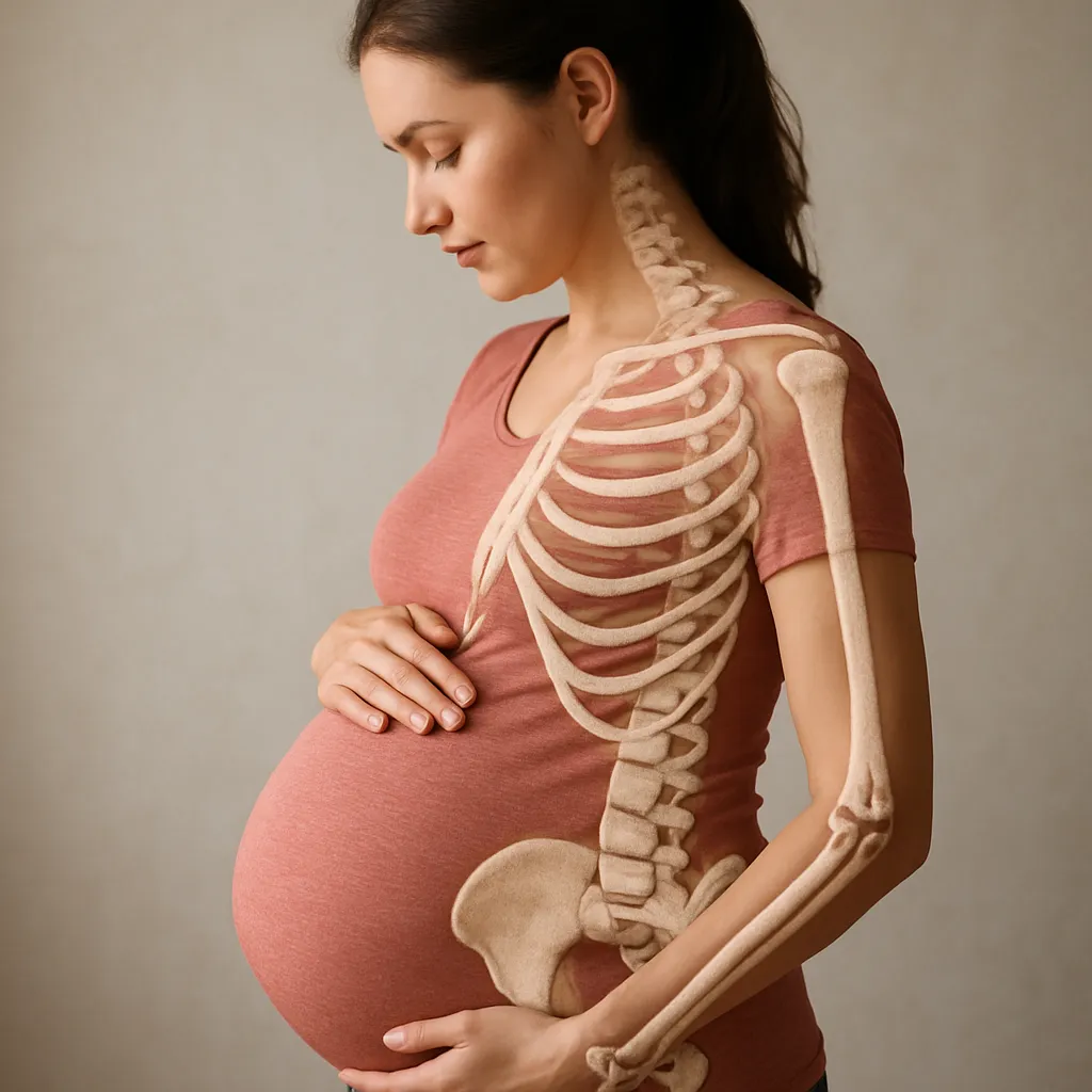
During pregnancy, a woman’s body undergoes profound physiological transformations to support the growing fetus. Among these adaptations, changes in bone density attract significant attention from researchers and clinicians. Understanding how pregnancy influences skeletal health is essential for developing preventive strategies and ensuring optimal outcomes for both mother and child. This article explores the multifaceted effects of gestation on maternal bone, highlights nutritional and hormonal factors, examines postpartum dynamics, and outlines long-term considerations.
Physiological Changes in Bone During Pregnancy
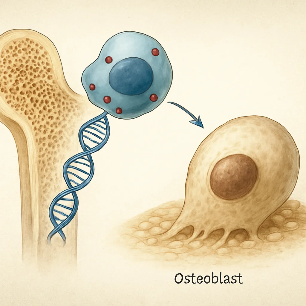
Bone is an active tissue, constantly undergoing remodeling to adapt to mechanical stresses and metabolic demands. During gestation, shifts in blood volume, weight distribution, and endocrine signaling converge to alter bone turnover rates. Elevated levels of placenta-derived hormones such as estrogen, progesterone, and relaxin modulate osteoclast and osteoblast activity. Early in pregnancy, estrogen promotes bone formation, while later phases see a relative increase in resorption to release minerals needed for fetal development.
Mechanical loading also shifts as the uterus expands, changing posture and center of gravity. These biomechanical adjustments can stimulate localized remodeling in the lumbar spine and pelvis. Studies using dual-energy X-ray absorptiometry (DEXA) report modest, transient declines in bone mass in specific skeletal sites, especially during the third trimester. However, comprehensive data suggest that many women experience recovery of bone mass within months postpartum, indicating a dynamic but reversible process.
At the cellular level, pregnancy influences the balance between bone formation and resorption in favor of maintaining calcium homeostasis for the fetus. Enhanced intestinal absorption and renal conservation of minerals support this balance, but bone still serves as a critical reservoir. The interplay between mechanical cues and endocrine signals underscores the complexity of skeletal adaptation during gestation.
Role of Nutrition and Calcium Homeostasis
Proper nutrition is a cornerstone of preserving maternal bone health. The fetus requires significant amounts of calcium and phosphate for skeletal development, particularly in the third trimester when mineral accretion peaks. To meet these demands, the mother’s body enhances intestinal absorption of dietary calcium via increased active transport mechanisms mediated by vitamin D. Meanwhile, renal excretion of calcium diminishes, conserving crucial stores.
When dietary intake is insufficient, the maternal skeleton may partially fulfill fetal requirements, leading to temporary bone loss. To minimize this effect, recommendations emphasize a balanced diet rich in calcium sources:
- Dairy products (milk, cheese, yogurt)
- Leafy green vegetables (kale, spinach)
- Fortified plant-based beverages
- Fish with edible bones (sardines, salmon)
- Nuts and seeds (almonds, chia seeds)
Besides calcium, adequate protein, magnesium, vitamin K, and trace elements support proper bone mineralization. Omega-3 fatty acids may also play a role in reducing inflammatory cytokines that promote bone resorption. Supplementation strategies should be tailored to individual risk profiles, with healthcare providers monitoring serum calcium and 25-hydroxyvitamin D levels throughout pregnancy.
Additionally, the exchange of minerals between mother and fetal skeleton follows a graded mechanism: the placenta actively transports calcium to the fetus, ensuring that even in cases of borderline maternal status, fetal needs take precedence. This prioritization demonstrates the evolutionary importance of skeletal development but also underscores the potential vulnerability of the maternal bone bank.
Postpartum and Lactation Impact
After childbirth, rapid shifts in hormone levels trigger further adaptations. Declining estrogen and progesterone levels may transiently promote bone resorption until menstrual cycles resume. Simultaneously, if a mother chooses to breastfeed, additional skeletal changes occur to supply calcium in breast milk. During lactation, daily calcium losses can reach 200–300 mg, prompting accelerated bone turnover.
Bone resorption during lactation is mediated by elevated levels of prolactin and parathyroid hormone-related peptide (PTHrP). However, this bone loss is typically reversible once weaning occurs. Longitudinal studies reveal that most women recover pre-pregnancy bone mass within 6–12 months after stopping breastfeeding, reflecting the plasticity of the maternal skeleton.
Factors influencing postpartum bone dynamics include the duration of breastfeeding, spacing between pregnancies, baseline nutritional status, and engagement in weight-bearing exercises. Weight-bearing activities and resistance training during the postpartum period can help stimulate osteoblastic activity and promote recovery of bone mass. Healthcare professionals often recommend tailored exercise programs to support both pelvic floor rehabilitation and skeletal health.
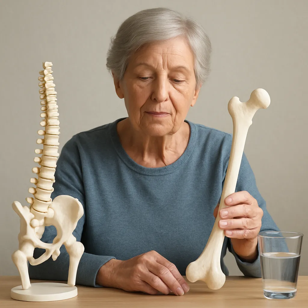
Long-term Consequences and Prevention Strategies
While temporary bone loss during gestation and lactation is expected, long-term consequences vary among individuals. Women with preexisting low bone mass, nutritional deficiencies, or multiple closely spaced pregnancies may face an elevated risk of developing osteoporosis later in life. Conversely, pregnancy can serve as a catalyst for beneficial lifestyle changes, encouraging improved nutrition and physical activity.
Preventive strategies for safeguarding maternal skeletal health encompass several key approaches:
- Ensuring adequate daily intake of calcium (1,000–1,300 mg) and vitamin D (600–800 IU) through diet and supplements
- Engaging in regular weight-bearing and resistance exercises to stimulate bone formation
- Monitoring bone density in high-risk individuals via DEXA scanning
- Spacing pregnancies to allow for full skeletal recovery between lactation periods
- Addressing lifestyle factors such as smoking cessation and moderate alcohol consumption
Early identification of at-risk mothers allows for targeted intervention during the reproductive years, optimizing peak bone mass before menopausal bone loss begins. Collaborative care between obstetricians, endocrinologists, nutritionists, and physiotherapists can foster a multidisciplinary framework to support women across the perinatal period.
Ultimately, pregnancy is a time of remarkable physiological flexibility. By understanding the mechanisms of skeletal adaptation and implementing evidence-based preventive measures, clinicians can empower women to navigate these changes while preserving long-term bone health.