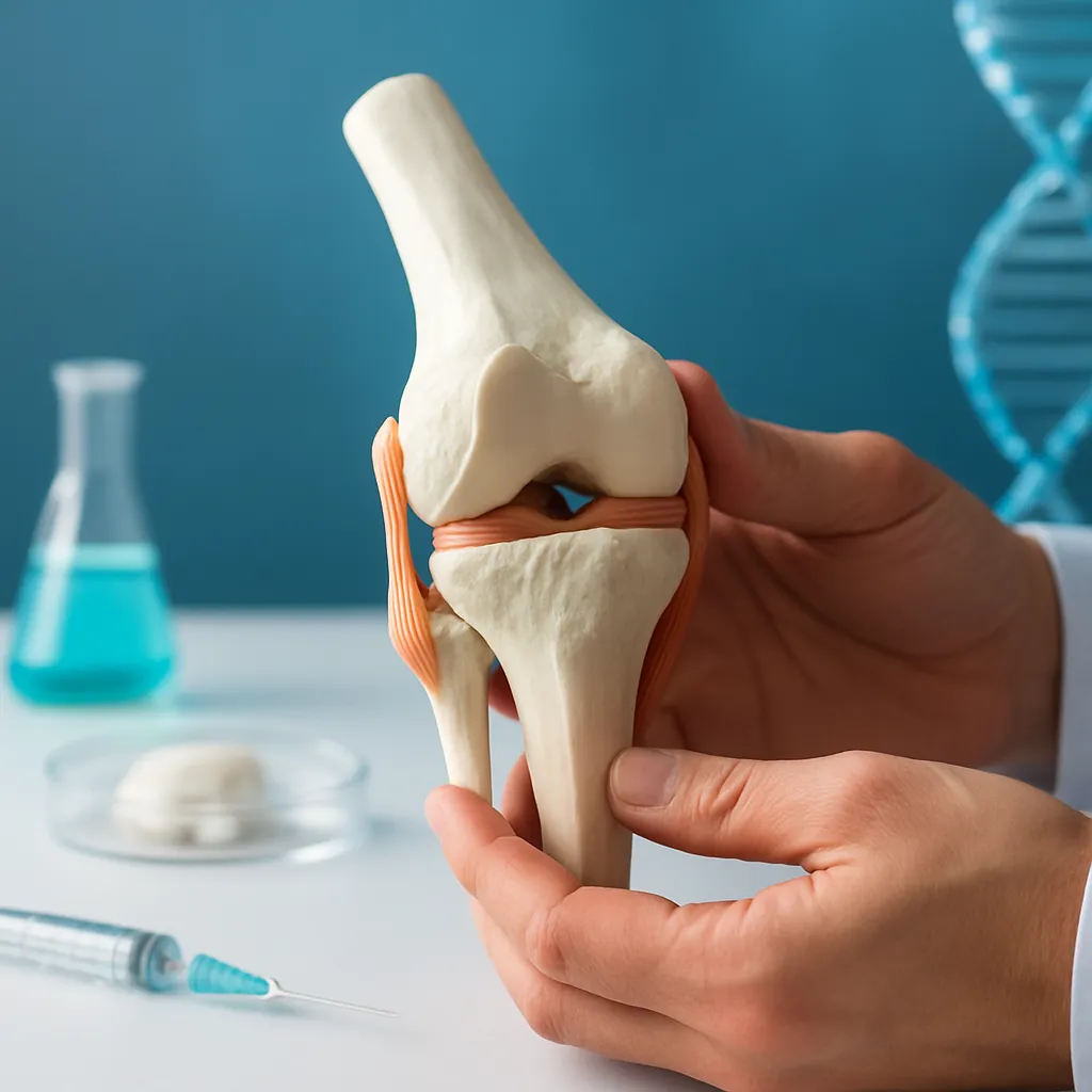




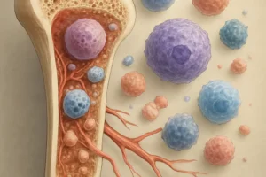
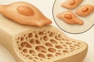


Bone Morphogenetic Proteins (BMPs) have revolutionized the field of orthopedics by harnessing the body’s intrinsic ability to direct cellular differentiation and tissue formation. Initially identified as critical regulators of **osteogenesis**, BMPs are now at the forefront of bioengineering strategies to enhance bone **regeneration**, fusion, and repair. This article explores BMP biology, **clinical** applications, and emerging challenges in the design of next-generation therapies for musculoskeletal disorders.
Historical Perspective and Molecular Foundations
The discovery of BMPs in the 1960s provided a paradigm shift in understanding how bone tissue forms. Through in vivo assays that induced ectopic bone in muscle tissue, researchers uncovered a family of signaling molecules belonging to the Transforming Growth Factor-β superfamily. Key members such as BMP-2, BMP-7, and BMP-4 coordinate intricate cellular programs that govern proliferation, migration, and differentiation of mesenchymal stem cells (MSCs) into osteoblasts and chondrocytes.
Mechanism of Action
Upon binding to type I and type II BMP receptors on target cells, BMPs trigger receptor serine/threonine kinase activity, leading to phosphorylation of Smad proteins (Smad1/5/8). These phosphorylated Smads complex with Smad4 and translocate into the nucleus to modulate transcription of osteogenic genes such as Runx2, Osterix, and alkaline phosphatase. Through feedback loops involving **cytokines**, inhibitors like Noggin or SMURF proteins tightly regulate BMP signaling to prevent aberrant bone formation.
Recombinant Technologies
- Production of rhBMP-2 and rhBMP-7 via bacterial or mammalian expression systems
- Purification strategies ensuring **biocompatibility** and bioactivity
- Formulation with carrier matrices (collagen sponges, hydrogels, ceramics)
Clinical Applications in Orthopedic Surgery
The translation of BMPs into **clinical** practice has centered on spinal fusion, trauma, and complex bone defects. The United States Food and Drug Administration (FDA) approved rhBMP-2 for anterior lumbar interbody fusion and rhBMP-7 for nonunion fractures, ushering in a new era of growth-factor augmented repairs.
Spinal Fusion Procedures
Spinal arthrodesis requires stable and durable intervertebral fusion. Incorporation of rhBMP-2 with a collagen-based scaffold has demonstrated higher fusion rates compared to autograft alone, reducing donor-site morbidity and shortening recovery times. Nevertheless, BMP-induced ectopic bone formation and postoperative inflammation remain areas of ongoing research.
Nonunion Fractures and Pseudarthrosis
Persistent nonunion represents a significant challenge in orthopedic trauma. Use of rhBMP-7 combined with autograft or **allograft** has shown robust stimulation of bone **healing**, even in patients with compromised regenerative potential due to comorbidities such as diabetes or smoking. Controlled clinical trials report union rates exceeding 80% when BMPs are employed adjunctively.
Large Bone Defect Reconstruction
Critical-size defects following tumor resection or high-energy trauma often exceed the body’s native reparative capacity. BMPs, delivered via **biomaterials** scaffolds—ranging from calcium phosphate ceramics to biodegradable polymers—support new bone deposition and scaffold resorption in a synchronized manner. Emerging designs incorporate nanofibrous architectures and controlled release kinetics for sustained BMP bioavailability.
Advanced Delivery Systems and Biomaterial Innovations
Successful BMP therapy hinges on achieving a precise spatial and temporal presentation of growth factors. Recent advances focus on multifunctional scaffolds that integrate mechanical support with biological cues.
Hydrogel-Based Carriers
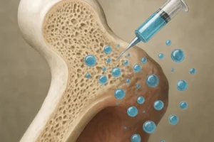
Injectable hydrogels composed of hyaluronic acid, polyethylene glycol (PEG), or modified chitosan can encapsulate BMPs and maintain local **bioactivity**. Crosslinking density and degradation rate are tailored to match the healing timeline, ensuring gradual BMP release and minimizing burst effects.
Nanoparticle–BMP Conjugates
- PLGA (poly(lactic-co-glycolic acid)) nanoparticles decorated with BMP-2 for enhanced cellular uptake.
- Gold and silica nanoparticles serving as carriers and imaging agents for real-time tracking.
- Stimuli-responsive systems (pH, enzyme, or temperature sensitive) for on-demand BMP liberation.
Composite Scaffolds
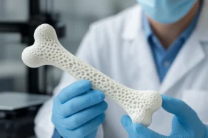
New composite scaffolds physically mimic trabecular bone architecture. By combining hydroxyapatite granules with polymeric matrices, these constructs provide **osteoconductive** surfaces while releasing BMPs from embedded microspheres. Mechanical properties approximate those of cancellous bone, facilitating load transfer and preventing stress shielding.
Safety Considerations and Regulatory Landscape
Despite significant clinical successes, BMP therapies carry potential risks. Dose-related complications include inflammatory responses, seroma formation, and heterotopic ossification. Careful patient selection and optimized delivery platforms are essential to mitigate adverse outcomes.
Immunogenicity and Dose Optimization
High local concentrations of BMP may trigger immune cell infiltration and increased production of inflammatory mediators. Dose-ranging studies aim to identify the minimal effective concentration that yields reliable bone formation without systemic spillover.
Regulatory Challenges
- Validation of Good Manufacturing Practice (GMP) for consistent rhBMP production
- Preclinical models to predict long-term safety, including carcinogenesis risk
- Post-market surveillance to assess real-world efficacy and rare complications
Emerging Trends and Future Directions
Continuous innovation is expanding the therapeutic scope of BMPs. Combinatorial approaches merging BMPs with angiogenic factors (VEGF, FGF) aim to synchronize vascular ingrowth and bone tissue formation. Gene therapy strategies use viral and non-viral vectors to deliver BMP genes directly to defect sites, achieving sustained local production by host cells.
Personalized Regenerative Medicine
Advances in 3D printing enable patient-specific implants impregnated with BMP-loaded microspheres. Collaboration between orthopedic surgeons, bioengineers, and data scientists is steering toward tailored graft solutions that account for defect geometry, mechanical loading, and individual healing capacity.
Stem Cell–BMP Synergies
Combining MSCs with BMPs within three-dimensional niches shows promise in preclinical models. MSCs not only serve as progenitors for osteoblasts but also secrete a milieu of **cytokines** and matrix proteins that facilitate engraftment and new tissue maturation. Bioreactor systems for cell-scaffold conditioning prior to implantation are under active investigation.