



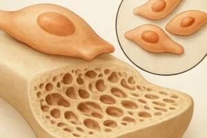


Bone is a dynamic tissue that continuously undergoes structural renewal to maintain strength and integrity. The interplay between cellular actors, biochemical signals, and external forces ensures an optimal balance between formation and resorption. Understanding how mechanical cues influence the bone’s internal machinery provides critical insight into health, disease, and therapeutic innovation.
Bone Remodeling: Cellular and Molecular Foundations
Bone remodeling is orchestrated by a sophisticated network of cells and signals. The triad of osteoclasts, osteoblasts, and osteocytes work in concert to remove old or damaged matrix and deposit new mineralized tissue. This cycle can be divided into four overlapping phases: activation, resorption, reversal, and formation.
Activation begins when lining cells retract and expose the bone surface. Local factors such as cytokines and growth factors—including RANKL and OPG—govern the differentiation of mononuclear precursors into osteoclasts, which adhere to bone and secrete acids and proteases to dissolve mineral and collagen. Following a controlled resorption period, macrophage-like reversal cells prepare the surface for new matrix deposition.
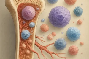
In the formation phase, mesenchymal stem cells differentiate into osteoblasts, synthesizing collagen type I and non-collagenous proteins. Mineralization follows, yielding rigid lamellar bone. A fraction of osteoblasts undergoes apoptosis, some become lining cells, and others embed in the matrix to differentiate into osteocytes. These embedded cells form an extensive lacuno-canalicular network for nutrient transport and signal transduction.
The precise regulation of remodeling is mediated by systemic hormones—such as parathyroid hormone (PTH), calcitonin, and sex steroids—and local factors, including transforming growth factor-β (TGF-β) and bone morphogenetic proteins (BMPs). Disruption of this equilibrium can lead to skeletal disorders, highlighting the importance of both cellular function and biochemical milieu.
Mechanotransduction: How Mechanical Forces Shape Bone Tissue
Mechanical loading is a potent regulator of bone mass and geometry. Cells within bone can sense and respond to mechanical stimuli through a process called mechanotransduction. When mechanical forces such as compression, tension, or fluid shear stress are applied to bone, they generate microstrain within the matrix, prompting a cascade of biochemical events.
Osteocytes, the primary mechanosensors in bone, detect deformation via their dendritic processes. Fluid flow through canaliculi produces shear stress that activates ion channels and second-messenger systems. Key pathways include the activation of integrins, focal adhesion kinase (FAK), and subsequent phosphorylation of proteins involved in gene expression. The Piezo channels on osteocyte membranes serve as stretch-activated channels, allowing rapid Ca2+ influx and initiation of downstream signals.
Key Pathways in Mechanosensing
- Wnt/β-catenin signaling: Mechanical load upregulates Wnt ligands, promoting osteoblastogenesis and inhibiting apoptosis of bone-forming cells.
- IGF-1 production: Shear stress elevates insulin-like growth factor 1, enhancing matrix synthesis and cell proliferation.
- Nitric oxide (NO) release: NO acts as a paracrine factor to regulate osteoclast activity and vasodilation, improving nutrient supply.
- Prostaglandins (PGE2): Synthesized rapidly in response to stretch, they modulate gene expression related to bone remodeling.
The magnitude, frequency, and duration of mechanical stimuli determine the nature of the bone response. Low-magnitude, high-frequency vibrations can stimulate bone formation with minimal energy input. Conversely, excessive or abnormal loading patterns may induce microdamage, invoking targeted resorptive activity to preserve structural integrity. Through these finely tuned responses, bone adapts its architecture—modifying thickness, porosity, and shape—to meet functional demands.
Clinical Implications and Future Directions
Understanding the synergy between mechanical forces and bone cell biology has profound clinical implications. Age-related bone loss and diseases such as osteoporosis reflect an imbalance favoring resorption over formation. Tailored exercise regimens, including resistance training and weight-bearing activities, harness the principles of mechanotransduction to enhance bone density and adaptation. Rehabilitation protocols for fracture healing often incorporate controlled mechanical loading to stimulate callus formation and optimize recovery.
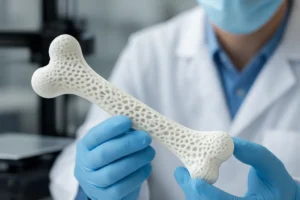
Orthopedic implants also benefit from insights into mechanical regulation. Surface topography and material stiffness influence peri-implant bone integration. Designing biomimetic scaffolds that replicate the native microarchitecture and mechanical properties of bone promotes rapid osseointegration and long-term stability. Advances in 3D printing enable patient-specific implants that distribute stress uniformly, reducing the risk of stress shielding and implant loosening.
Spaceflight research highlights another dimension: skeletal unloading in microgravity leads to rapid bone loss. Countermeasures include resistive exercise devices and pharmacological interventions targeting signaling pathways involved in mechanosensation. These studies not only safeguard astronaut health but also inform strategies for immobilized patients on Earth.
Future directions in bone medicine emphasize regenerative approaches, such as stem cell therapies and gene editing to enhance anabolic pathways. Combining mechanical cues with targeted delivery of growth factors may unlock new potentials for skeletal repair. Integrating computational models of bone mechanics with real-time monitoring technologies promises personalized interventions that optimize bone health throughout life.