




Effective recovery following orthopaedic procedures relies heavily on the intricate process of bone remodeling. This dynamic interplay between bone resorption and formation ensures not only structural integrity but also long-term functional restoration. Understanding these mechanisms provides clinicians and researchers with insights to optimize postoperative care and develop innovative therapies.
Biological Mechanisms of Bone Remodeling
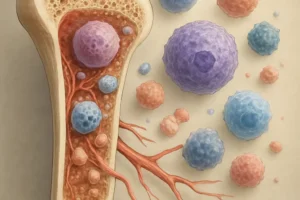
Bone remodeling is orchestrated by specialized cells and signaling molecules that maintain skeletal homeostasis. The key players include osteoclasts, responsible for bone resorption, and osteoblasts, which drive new bone formation. This cyclical process comprises precise phases that overlap and depend on each other for complete repair.
Stage 1: Inflammation and Cell Recruitment
Immediately after surgical intervention, a controlled inflammatory response occurs. Damaged tissues release cytokines and growth factors that attract precursor cells to the site. Vascular disruption triggers acute hemostasis followed by angiogenesis, providing essential oxygen and nutrients. Key events include:
- Platelet degranulation and fibrin clot formation
- Secretion of interleukins (IL-1, IL-6) and tumor necrosis factor (TNF-α)
- Recruitment of mesenchymal stem cells (MSCs)
Stage 2: Bone Resorption
Osteoclasts differentiate from monocyte/macrophage lineages under the influence of receptor activator of nuclear factor κB ligand (RANKL). These multinucleated cells adhere to the bone matrix and secrete hydrochloric acid and proteolytic enzymes, creating resorption pits. This controlled breakdown removes microdamage and prepares the surface for new bone deposition.
Stage 3: Bone Formation
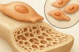
Following resorption, osteoblasts migrate to the cleared areas. They synthesize collagen type I and non-collagenous proteins, forming an organic osteoid matrix. Mineralization then occurs through deposition of hydroxyapatite crystals, restoring mechanical strength. Balancing resorption and formation is critical; any disruption can lead to delayed healing or pathological bone loss.
Clinical Implications After Surgical Intervention
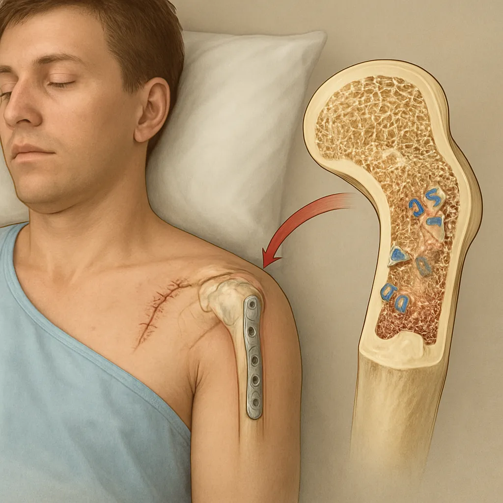
Surgeries such as joint arthroplasty, spinal fusion, and fracture fixation impose unique challenges to bone remodeling. Implant design, surgical technique, and local biological environment dictate the healing trajectory. Optimizing these factors can dramatically improve outcomes.
Role of Biomaterials in Healing
Advances in biomaterials have introduced scaffolds, coatings, and growth factor delivery systems that aim to enhance cellular responses. Examples include:
- Porous titanium implants that promote osteointegration through mechanical interlocking
- Calcium phosphate cements for bone void filling and gradual resorption
- Collagen-hydroxyapatite composites delivering recombinant bone morphogenetic proteins (BMPs)
Mechanical Stabilization and Mechanotransduction
Proper load distribution and stability are crucial for guiding cellular activity. Mechanotransduction pathways sense mechanical stimuli and convert them into biochemical signals, promoting osteogenesis. Rigid fixation devices and adjustable external frames help maintain optimal strain levels to avoid stress shielding and ensure consistent remodeling.
Factors Influencing Successful Remodeling
Multiple internal and external factors can accelerate or impede bone repair. Addressing each element holistically is key to achieving robust, long-lasting results.
- Nutrition: Adequate intake of calcium, vitamin D, protein, and trace minerals supports matrix synthesis and mineralization.
- Hormonal milieu: Estrogen, parathyroid hormone, and growth hormone regulate osteoblast and osteoclast activity.
- Age and comorbidities: Elderly patients and those with osteoporosis or diabetes often exhibit slower remodeling rates.
- Pharmacological agents: Bisphosphonates, selective estrogen receptor modulators (SERMs), and anabolic therapies can modulate remodeling dynamics.
- Smoking and lifestyle: Tobacco use and sedentary habits impair vascularization and cellular proliferation.
Advanced Imaging and Monitoring Techniques
Accurate assessment of bone healing is essential for timely interventions. Modern imaging modalities allow for noninvasive evaluation of both structure and metabolic activity.
Dual-Energy X-Ray Absorptiometry (DXA)
DXA provides quantitative measurement of bone mineral density (BMD). Serial scans can track changes during the postoperative period, indicating successful integration or detecting risks of implant loosening.
Computed Tomography (CT) and Micro-CT
CT imaging offers high-resolution anatomical detail. Micro-CT, primarily used in research, quantifies trabecular architecture and porosity, revealing microstructural remodeling patterns.
Magnetic Resonance Imaging (MRI) and Positron Emission Tomography (PET)
MRI excels at visualizing soft tissue and marrow changes, while PET with tracers like fluorodeoxyglucose (FDG) can highlight regions of high metabolic activity. Combining these modalities enhances early detection of complications.
Emerging Therapeutic Strategies
Ongoing research focuses on harnessing biological pathways to accelerate regeneration. Innovations include cell-based therapies, gene editing, and targeted molecular treatments.
- MSC transplantation to provide osteoprogenitor cells directly at the injury site
- CRISPR/Cas9 gene modulation of osteogenic factors to enhance local bone formation
- Nanoparticle-based delivery of peptides and microRNAs to fine-tune cellular signaling
- Smart implant coatings that release anti-inflammatory agents and antibiotics on demand
By integrating insights from cellular biology, materials science, and biomechanics, the future of postoperative bone remodeling promises more predictable healing pathways, reduced complications, and improved patient quality of life.