




The intricate process of bone mineralization forms the cornerstone of skeletal integrity, facilitating mobility and protecting vital organs. This article delves into the cellular, molecular, and hormonal mechanisms that govern this dynamic equilibrium. By exploring the roles of bone-forming cells, key regulators such as parathyroid hormone and vitamin D, and the impact of nutritional and therapeutic interventions, we illuminate pathways toward maintaining optimal bone health and combating disorders like osteoporosis.
Mechanisms of Bone Mineralization
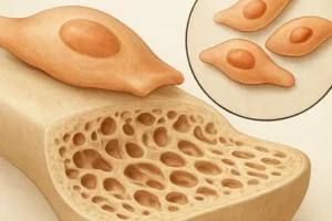
Bone mineralization is a highly coordinated process driven by specialized cells and extracellular matrix components. Osteoblasts secrete collagen type I, forming an organic scaffold called osteoid. Mineral deposition occurs when calcium and phosphate ions crystallize as hydroxyapatite on this matrix. The following steps are crucial:
- Matrix Production: Osteoblasts produce collagen fibrils and non-collagenous proteins such as osteocalcin and osteopontin.
- Vesicle Release: Matrix vesicles enriched in alkaline phosphatase concentrate calcium and phosphate.
- Crystal Nucleation: Supersaturation of ions triggers hydroxyapatite nucleation within vesicles.
- Crystal Growth: Hydroxyapatite crystals propagate along collagen fibers, strengthening the bone matrix.
Coordinate interplay between osteoblasts, osteoclasts, and osteocytes ensures continual renewal through bone remodeling. Osteocytes, embedded in lacunae, sense mechanical strain and orchestrate local mineralization or resorption by signaling to surface cells.
Hormonal Regulation of Bone Metabolism
The endocrine system exerts profound influence on bone turnover. Key hormones modulate cellular activities:
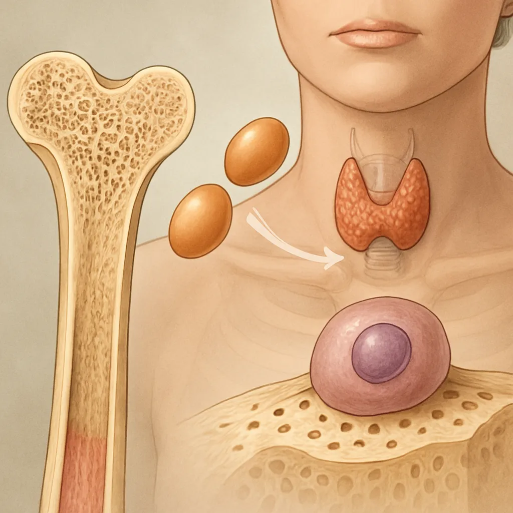
- Parathyroid Hormone (PTH): Secreted in response to low serum calcium, PTH stimulates osteoclastogenesis via RANKL expression on osteoblasts, enhancing bone resorption and calcium release.
- Calcitonin: Produced by thyroid C cells, it inhibits osteoclast function, reducing resorption and favoring mineral deposition.
- Vitamin D: Converted to calcitriol in the kidney, it increases intestinal calcium absorption and works synergistically with PTH to maintain mineral homeostasis.
- Sex Steroids (Estrogen and Testosterone): These hormones promote osteoblast activity and suppress osteoclastogenesis. Estrogen upregulates osteoprotegerin, a decoy receptor for RANKL, protecting against excessive resorption.
- Growth Hormone and IGF-1: Stimulate osteoblast proliferation and collagen synthesis, enhancing peak bone mass during adolescence.
- Glucocorticoids: Chronic excess impairs osteoblast differentiation and induces apoptosis, leading to bone loss.
Imbalances in these factors can tip the remodeling scale toward net bone loss, setting the stage for metabolic bone diseases.
Nutritional and Environmental Influences
Optimal bone mineralization relies on adequate nutrients and lifestyle factors:
- Calcium and Phosphate Intake: Dietary sources such as dairy, leafy greens, and fortified products supply essential minerals.
- Vitamin D Sufficiency: Sunlight-driven synthesis and supplementation ensure proper absorption of calcium.
- Protein Intake: Supports collagen matrix formation but requires balance to prevent acid load that may leach calcium.
- Physical Activity: Mechanical loading stimulates osteocyte signaling and promotes bone formation, enhancing density.
- Smoking and Alcohol: Both are detrimental, impairing osteoblast function and increasing resorption risk.
- Microbiome Interactions: Emerging evidence links gut flora to mineral absorption and systemic inflammation, impacting bone health.
Public health measures focusing on diet, exercise, and avoidance of modifiable risks can bolster skeletal resilience throughout life.
Clinical Implications and Therapeutic Approaches
Understanding bone mineralization and hormonal control has fueled advances in treating metabolic bone disorders. Current strategies include:
- Bisphosphonates: Analogues of pyrophosphate that inhibit osteoclast-mediated resorption by inducing apoptosis.
- Denosumab: A monoclonal antibody targeting RANKL, effectively reducing osteoclast activity and increasing bone mass.
- Selective Estrogen Receptor Modulators (SERMs): Mimic estrogen’s protective effects on bone without adverse risks in breast and uterine tissues.
- Teriparatide: Recombinant PTH fragment administered intermittently to stimulate osteoblasts and bone formation.
- Romosozumab: A sclerostin inhibitor that enhances Wnt signaling, promoting osteoblast-driven bone formation.
Emerging therapies targeting molecular pathways such as Wnt signaling and the endocannabinoid system promise further refinements. Precision medicine approaches, integrating genetic profiling and biomarker monitoring, aim to tailor treatments and optimize outcomes for individuals at risk of fractures and debilitating bone loss.