



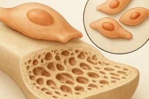

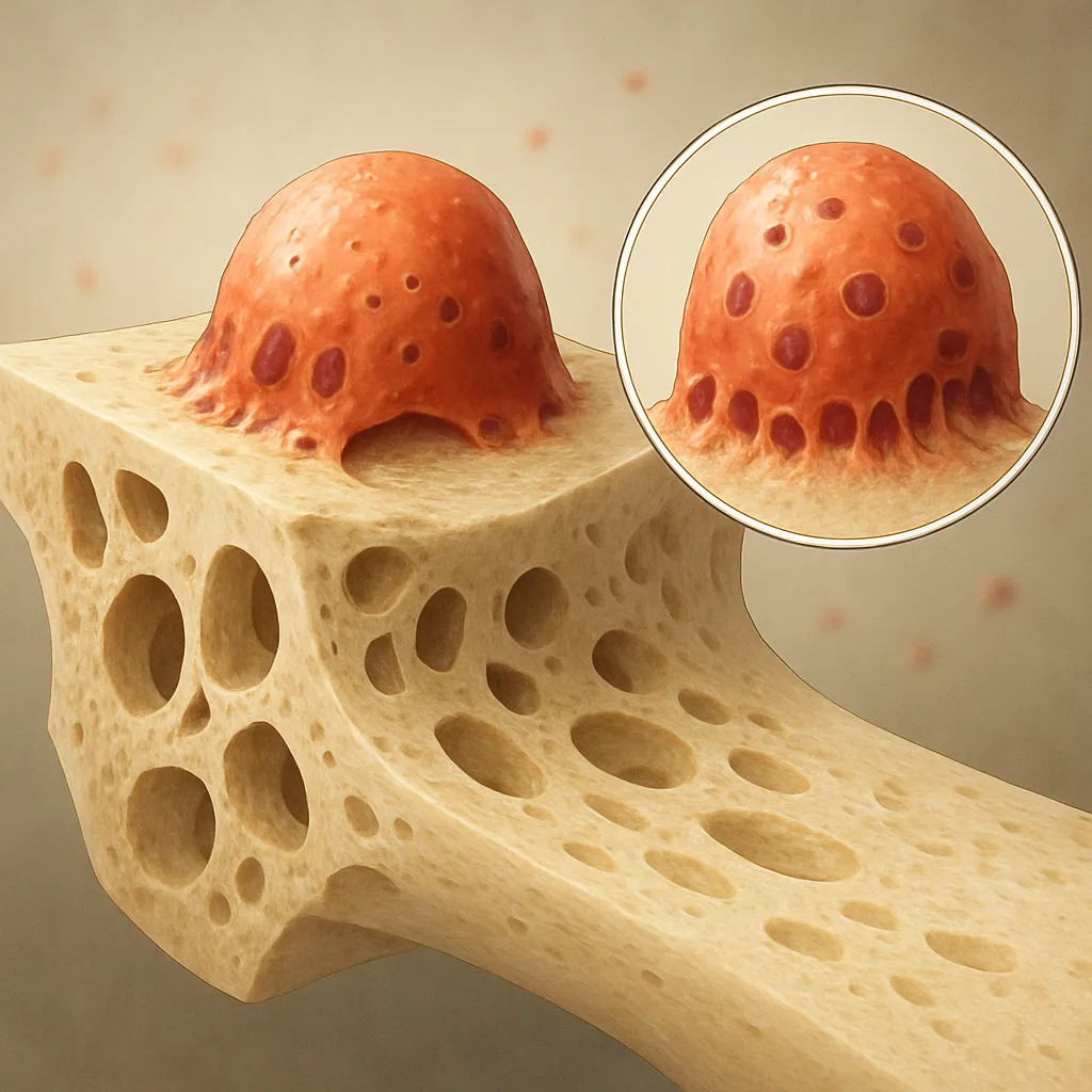
The dynamic equilibrium of the skeletal system relies on a finely tuned interplay between bone formation and bone resorption. At the heart of this process are osteoclasts, specialized multinucleated cells responsible for breaking down mineralized matrix. By examining the molecular signaling pathways, cellular interactions, and clinical relevance of these cells, we can deepen our understanding of bone health and disease. This article explores the essential roles of osteoclasts in bone turnover, the mechanisms guiding their differentiation, and the impact of dysregulation on disorders such as osteoporosis.
Bone Remodeling: An Overview
Bone remodeling is a lifelong process that ensures the maintenance of skeletal integrity, adaptation to mechanical stress, and mineral homeostasis. The cycle consists of four overlapping phases: activation, resorption, reversal, and formation. In the resorption phase, osteoclasts attach to the bone surface, form a specialized sealing zone, and secrete acidic and proteolytic enzymes to degrade the organic and inorganic matrix. This resorptive activity creates microscopic cavities that are subsequently replenished by osteoblasts during the formation phase.
Activation Phase and Cellular Recruitment
Initiation of remodeling begins with microdamage or systemic signals such as hormonal fluctuations. Osteocytes, the most abundant bone cells, sense mechanical strain and release factors like RANKL (Receptor Activator of Nuclear Factor κB Ligand), which is pivotal for osteoclastogenesis. The balance between RANKL and its decoy receptor, osteoprotegerin (OPG), determines the rate of osteoclast formation. Elevated RANKL or reduced OPG levels shift the equilibrium toward increased bone resorption.
Resorption by Osteoclasts
Once recruited, osteoclasts polarize and form the ruffled border, a membrane specialization that maximizes the surface area for matrix degradation. Through the secretion of hydrochloric acid (mediated by vacuolar H+-ATPases) and proteases such as cathepsin K, the inorganic hydroxyapatite crystals and organic collagen network are dissolved. The released calcium and phosphate enter the bloodstream, contributing to mineral homeostasis.
Cellular Mechanisms of Osteoclast Function
At the molecular level, osteoclast differentiation and activity depend on precise signaling events and interactions with other bone cells. Understanding these mechanisms provides insights into potential targets for therapeutic intervention in bone diseases.
Signaling Pathways Governing Differentiation
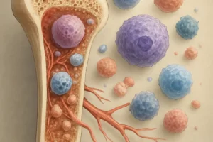
Osteoclast precursors originate from hematopoietic stem cells in the bone marrow. Two key cytokines, macrophage colony-stimulating factor (M-CSF) and RANKL, are indispensable for the progression of mononuclear precursors into mature multinucleated osteoclasts. The binding of RANKL to its receptor RANK on precursor cells triggers the recruitment of adaptor proteins like TRAF6 and activation of NF-κB, NFATc1, and AP-1 transcription factors. These transcriptional regulators orchestrate the expression of genes necessary for cell fusion, cytoskeletal organization, and resorptive machinery.
Role of Integrins and the Sealing Zone
The formation of the sealing zone is mediated by β3 integrins, which anchor osteoclasts to the bone surface. This adhesion complex, also known as the podosome belt, creates a microenvironment in which enzymes and protons act efficiently. Disruption of integrin signaling impairs bone resorption, demonstrating the critical role of cytoskeletal dynamics in osteoclast function.
Coupling of Resorption and Formation
Bone remodeling is not solely about degradation; it also involves coordination with osteoblasts to ensure proper formation of new matrix. Osteoclasts release coupling factors, such as sphingosine-1-phosphate and transforming growth factor-β (TGF-β), which recruit osteoprogenitor cells and stimulate osteoblast differentiation. This crosstalk highlights the interdisciplinary nature of skeletal biology, where resorptive and formative processes are tightly linked.
Clinical Implications and Therapeutic Strategies
Dysregulation of osteoclast activity leads to a spectrum of skeletal disorders. Excessive resorption contributes to osteoporosis and bone metastases, whereas insufficient activity can cause osteopetrosis. Targeting osteoclasts offers effective approaches to restore bone balance and prevent fractures.
Osteoporosis and Anti-resorptive Agents
Osteoporosis is characterized by decreased bone density and structural deterioration, often resulting from elevated osteoclast-mediated resorption relative to formation. First-line therapies include bisphosphonates, which bind to bone mineral and induce osteoclast apoptosis. Denosumab, a monoclonal antibody against RANKL, effectively reduces osteoclast numbers and activity. Emerging treatments involve cathepsin K inhibitors that specifically block collagen degradation at the resorption lacunae.
Bone Metastasis and Tumor-induced Osteolysis
Cancer cells that metastasize to bone secrete factors such as parathyroid hormone–related protein (PTHrP) to upregulate RANKL expression by stromal cells and osteoblasts. This creates a vicious cycle of osteolysis and tumor growth. Therapeutic strategies combine anti-resorptive agents with targeted cancer therapies to disrupt this interaction and alleviate skeletal-related events.
Future Directions in Osteoclast Research
Advances in imaging, genomics, and single-cell technologies are unveiling new regulators of osteoclast biology. The identification of microRNAs, long non-coding RNAs, and novel signaling mediators presents opportunities for precision medicine. Modulation of immune cell–osteoclast interactions also holds promise, particularly in inflammatory bone diseases where cytokine networks exacerbate resorption.
Key Terms:
- Osteoclasts
- Bone remodeling
- RANKL
- Bisphosphonates
- Cathepsin K
- Integrins
- Cytokines
- Osteoporosis
- Resorption
- Signaling