



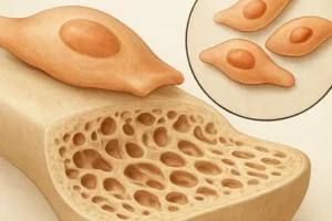


Bone health is governed by a tightly regulated interplay between formation and resorption processes, collectively known as bone remodeling. This dynamic equilibrium is crucial for maintaining skeletal integrity and mineral homeostasis. In chronic diseases, disruptions to this balance can lead to significant morbidity, characterized by reduced bone density, increased fracture risk, and altered microarchitecture. Understanding the underlying mechanisms of bone turnover, the available diagnostic markers, and the impact of various disease states is essential for developing effective therapeutic strategies.
Pathophysiology of Bone Turnover
Bone remodeling is orchestrated by two principal cell types: osteoblasts, responsible for new bone formation, and osteoclasts, which mediate bone resorption. These cells operate within the basic multicellular unit (BMU), where sequential activation, resorption, reversal, and formation phases ensure continuous renewal. The local and systemic regulators of this process include cytokines, hormones, and mechanical signals that together determine net bone balance.
Cellular Mechanisms
- Osteoclast differentiation is driven by receptor activator of nuclear factor κB ligand (RANKL) and macrophage colony-stimulating factor (M-CSF).
- Osteoblast activity depends on signaling pathways such as Wnt/β-catenin, bone morphogenetic proteins (BMPs), and insulin-like growth factor (IGF).
- The coupling of resorption and formation ensures that microdamage is repaired and mineral homeostasis is preserved.
Regulatory Factors
Systemic hormones—including parathyroid hormone (PTH), calcitonin, and sex steroids—interact with local mediators like interleukins (IL-1, IL-6) and tumor necrosis factor-alpha (TNF-α). Disruption in any of these pathways can shift the remodeling balance toward excessive resorption or inadequate formation, ultimately compromising bone strength.
Biochemical Markers and Diagnostic Tools
Evaluating bone turnover requires both imaging modalities and biochemical assays. These tools provide quantitative and qualitative insights into skeletal metabolism, enabling early detection of imbalance and monitoring of therapeutic efficacy.
Imaging Modalities
- Dual-energy X-ray absorptiometry (DXA) remains the gold standard for assessing bone mineral density (BMD).
- Quantitative computed tomography (QCT) offers volumetric density measurements and separates trabecular from cortical compartments.
- High-resolution peripheral quantitative computed tomography (HR-pQCT) can visualize microarchitectural changes at peripheral sites.
Biochemical Biomarkers
- Formation markers: Procollagen type I N-terminal propeptide (P1NP), osteocalcin, bone alkaline phosphatase (bALP).
- Resorption markers: C-terminal telopeptide of type I collagen (CTX), N-terminal telopeptide (NTX), tartrate-resistant acid phosphatase 5b (TRACP-5b).
- Emerging markers include sclerostin and dickkopf-1 (DKK1), which modulate Wnt signaling and may predict fracture risk.
Combining imaging and biochemical tests improves diagnostic accuracy. Temporal changes in biomarkers can precede measurable BMD alterations, allowing for earlier intervention. However, pre-analytical variables—such as circadian rhythms, food intake, and renal function—must be controlled to ensure reliable results.
Impact of Chronic Diseases on Bone Remodeling
Chronic conditions often exert adverse effects on skeletal metabolism through inflammatory mediators, altered hormonal profiles, and direct tissue damage. A thorough evaluation of bone turnover in these diseases aids in identifying patients at risk for secondary osteoporosis and fracture.
Rheumatoid Arthritis
Rheumatoid arthritis (RA) features systemic inflammation driven by TNF-α, IL-1, and IL-6, which promote osteoclastogenesis and inhibit osteoblast function. Patients exhibit both periarticular bone erosions and generalized bone loss. Frequent use of glucocorticoids, while controlling disease activity, further exacerbates bone resorption and impairs formation.
- Elevated CTX and decreased P1NP correlate with disease severity.
- Early DXA screening is recommended, especially when cumulative glucocorticoid dose exceeds 5 grams of prednisone equivalent.
Chronic Kidney Disease–Mineral and Bone Disorder
In chronic kidney disease (CKD), impaired phosphate excretion and decreased calcitriol synthesis disrupt mineral homeostasis, leading to secondary hyperparathyroidism and osteitis fibrosa. Bone turnover can be high, low, or mixed, necessitating bone biopsy in ambiguous cases. Management focuses on phosphate binders, vitamin D analogs, and calcimimetics to restore balance.
Endocrine Disorders
- Hyperthyroidism accelerates bone turnover by upregulating RANKL expression, increasing fracture risk despite normal BMD in some cases.
- Hypogonadism reduces sex steroid levels, leading to diminished bone formation and early onset of osteoporosis in men and women.
- Cushing’s syndrome induces catabolic effects on bone by antagonizing Wnt signaling and enhancing osteoclast activity.
Therapeutic Approaches and Future Directions
Treatment of bone turnover abnormalities in chronic diseases aims to restore the remodeling equilibrium, reduce fracture incidence, and alleviate skeletal pain. Current and emerging therapies target both resorption and formation pathways.
Anti-Resorptive Agents
- Bisphosphonates bind to hydroxyapatite and facilitate osteoclast apoptosis, effectively reducing resorption.
- Denosumab, a monoclonal antibody against RANKL, offers a reversible inhibition of osteoclastogenesis with biannual administration.
- Selective estrogen receptor modulators (SERMs) mimic estrogen’s protective effects on bone without adverse uterine or breast stimulation.
Anabolic Treatments
- Parathyroid hormone (teriparatide) stimulates osteoblast proliferation and activity when given intermittently.
- Abaloparatide, a PTH-related peptide analog, exhibits reduced hypercalcemia risk while enhancing bone formation.
- Emerging sclerostin inhibitors, such as romosozumab, enhance Wnt signaling to increase bone mass and improve microarchitecture.
Personalized Medicine and Biomarker-Guided Therapy
Advances in genomics and proteomics are paving the way for individualized treatment plans. Biomarker panels that integrate bone turnover markers, genetic polymorphisms, and disease-specific risk factors may optimize therapeutic selection and timing. Machine learning algorithms applied to longitudinal data sets hold promise for predicting treatment response and fracture risk with high precision.
Future Research Directions
- Development of noninvasive imaging techniques capable of assessing bone quality at the cellular level.
- Identification of novel molecular targets to uncouple formation from resorption, thereby maximizing anabolic effects.
- Exploration of microbiome–bone axis interactions, given emerging evidence that gut flora influence mineral absorption and inflammation.