




The intricate structure of the human skeleton extends beyond mere strength and support. Bone quality encompasses not only bone mineral density but also the delicate interplay of architectural, material, and mechanical properties. Recent strides in imaging technology offer an unprecedented window into the inner realm of skeletal health, enabling clinicians and researchers to evaluate microstructural changes, detect early signs of degeneration, and guide personalized treatment strategies. This article explores the cutting-edge approaches in bone quality assessment, focusing on advanced imaging modalities, quantitative analytic techniques, and their clinical implications.
Advanced Imaging Modalities for Bone Quality Assessment
Assessing bone integrity has evolved from simplistic two-dimensional projections to multidimensional, high-resolution techniques. These modalities promise superior characterization of bone’s complex morphology and material composition.
High-Resolution Peripheral Quantitative Computed Tomography (HR-pQCT)
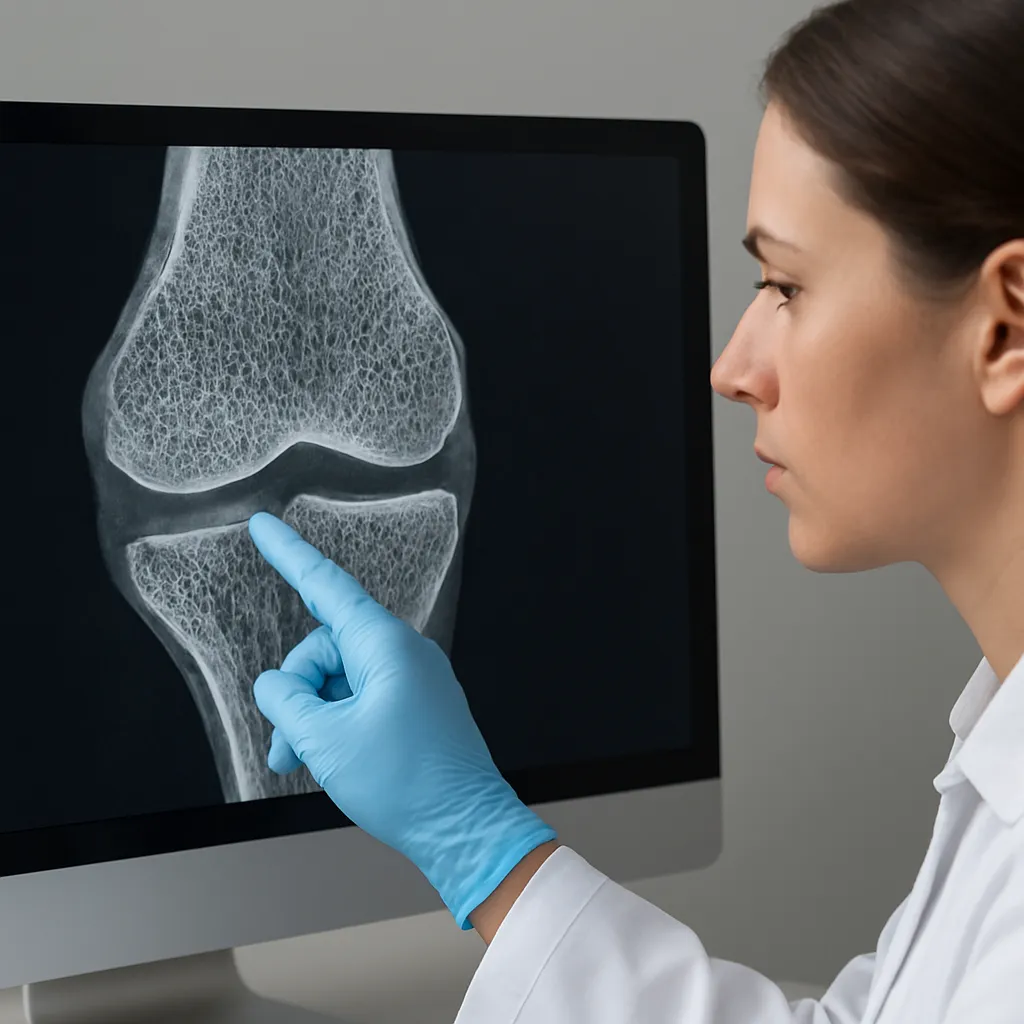
HR-pQCT represents a leap forward in visualizing both cortical bone and trabecular bone compartments at micrometer resolution. By acquiring volumetric scans of peripheral sites (distal radius and tibia), HR-pQCT enables:
- Separation of cortical porosity from cortical thickness
- Three-dimensional rendering of trabecular rods and plates
- Quantitative evaluation of bone microarchitecture
Studies demonstrate HR-pQCT’s capacity to predict fracture risk more accurately than conventional methods, as it captures alterations in microstructural parameters long before significant density loss occurs.
Magnetic Resonance Imaging (MRI) with Ultrashort Echo Time
Traditional MRI struggles to detect bone due to its rapid signal decay, yet the introduction of magnetic resonance imaging sequences with ultrashort echo times (UTE) overcomes this barrier. UTE-MRI facilitates:
- Visualization of cortical pores and bone water content
- Non-ionizing assessment of bone material properties
- Evaluation of collagen-bound water, reflecting bone toughness
By quantifying bound and free water in bone, UTE-MRI offers a non-invasive metric of matrix integrity, complementing density measures and illuminating early stages of disease such as osteoporosis.
Dual-Energy X-ray Absorptiometry (DXA) and Beyond
While dual-energy X-ray absorptiometry remains a clinical mainstay for bone mineral density (BMD) screening, its two-dimensional perspective limits insight into structural complexity. Innovations to enhance DXA include:
- Trabecular Bone Score (TBS): Texture analysis that infers trabecular microarchitecture.
- Vertebral Fracture Assessment (VFA): Low-dose lateral spine imaging for fracture detection.
These augmentations strive to remedy DXA’s inability to capture microstructural nuances, yet they still lag behind volumetric methods in spatial resolution.
Quantitative Metrics and Analytical Techniques
Beyond image acquisition, robust analytic frameworks are essential to translate visual data into actionable insights. Quantitative techniques range from simple geometric metrics to sophisticated computational models.
Microarchitectural Parameters
Key metrics derived from high-resolution datasets include:
- Bone Volume Fraction (BV/TV): Ratio of bone volume to total volume, indicating bone mass distribution.
- Trabecular Thickness (Tb.Th), Separation (Tb.Sp), and Number (Tb.N): Detailed descriptors of trabecular geometry.
- Cortical Porosity (Ct.Po): Proportion of void spaces within cortical bone, a critical determinant of strength.
These parameters can be mapped across skeletal sites to identify regions of compromised integrity and monitor therapeutic interventions.
Finite Element Analysis (FEA)
Finite element analysis translates imaging data into computational models that estimate mechanical behavior under physiological loads. FEA workflows involve:
- Segmentation of bone geometry from CT or HR-pQCT scans
- Assignment of material properties based on density–modulus relationships
- Simulation of loading scenarios (e.g., compression, bending, torsion)
The resulting maps of stress and strain pinpoint areas at high risk of microdamage, enhancing fracture prediction beyond density metrics alone.
Elastography and Mechanical Characterization
Emerging ultrasound-based elastography techniques measure the propagation of shear waves through bone, yielding shear modulus estimates that reflect material stiffness. Combined with high-frequency ultrasound, elastography offers a non-ionizing approach to assess bone elasticity and anisotropy.
Clinical Applications and Case Studies
The integration of advanced imaging into clinical practice has transformative implications for diagnosing and managing skeletal disorders.
Osteoporosis Management
Conventional diagnosis of osteoporosis relies on BMD thresholds, which may miss individuals with poor microarchitecture but near-normal density. By incorporating HR-pQCT and FEA-derived strength estimates:
- High-risk patients with compromised trabecular connectivity can be identified earlier.
- Treatment efficacy (e.g., bisphosphonates, denosumab) can be monitored through microstructural improvements rather than solely BMD changes.
Fracture Healing and Orthopedic Implants
Advanced imaging tracks the progression of callus formation and mineralization during fracture healing. Time-lapse HR-pQCT scans quantify:
- Callus volume and mineral density over healing phases
- Restoration of trabecular continuity in weight-bearing bones
In implantology, CT-based FEA predicts implant–bone interface stability, guiding implant design and surgical planning for improved osseointegration.
Metabolic Bone Diseases
Conditions such as osteogenesis imperfecta, Paget’s disease, and chronic kidney disease-related mineral bone disorder demand nuanced assessment of bone quality beyond density. Imaging biomarkers enable:
- Quantification of collagen cross-linking defects
- Monitoring of bone turnover rates via dynamic contrast-enhanced MRI
Challenges and Future Perspectives
Despite remarkable advancements, several challenges must be addressed to fully harness the potential of advanced imaging.
- Standardization: Variation in imaging protocols and analytic algorithms hampers cross-center comparisons and large-scale studies.
- Radiation Dose: CT-based methods involve ionizing radiation; minimizing dose while preserving resolution remains a priority.
- Cost and Accessibility: High-resolution modalities and computational modeling incur significant expense, limiting widespread clinical adoption.
Looking forward, integration of artificial intelligence for automated segmentation and predictive modeling promises to streamline workflows. Multimodal approaches combining imaging data with molecular biomarkers and genetic profiles could usher in a new era of precision orthopedics. By capturing the multifaceted nature of bone quality—spanning microarchitecture, material composition, and mechanical competence—these innovations pave the way for earlier diagnosis, personalized therapies, and improved skeletal health outcomes.