The human skeleton constantly adapts to the mechanical forces it encounters. Bone remodeling under mechanical stress is a finely tuned process that ensures structural integrity, mineral homeostasis, and functional performance. This dynamic interplay involves specialized cells, intricate signaling pathways, and the continuous reshaping of the bone matrix. Understanding these mechanisms provides valuable insights for orthopedics, sports medicine, and the treatment of metabolic bone disorders.
Physiological Basis of Bone Remodeling
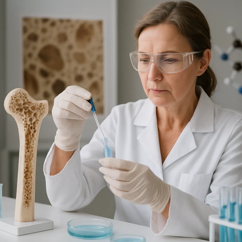
Bone is not a static tissue; it undergoes perpetual renewal through the coordinated actions of osteoblasts and osteoclasts. This balanced exchange maintains optimal strength and repairs microdamage generated by routine activity. Remodeling occurs in discrete units called basic multicellular units (BMUs), where bone resorption and formation occur sequentially. The remodeling cycle can be divided into the activation, resorption, reversal, formation, and mineralization phases. Each phase relies on precise communication between cells and their surrounding extracellular matrix, ensuring that bone mass and architecture are matched to mechanical demands.
Bone Cells and Their Roles
At the forefront of remodeling are three principal cell types:
- Osteoclasts: Multinucleated cells responsible for bone resorption. They dissolve mineral content and degrade organic matrix.
- Osteoblasts: Mononuclear cells that synthesize new matrix components, including type I collagen, and facilitate mineral deposition.
- Osteocytes: Former osteoblasts embedded within lacunae, acting as mechanosensors and regulators of the remodeling process.
Communication between these cells occurs via paracrine signals such as RANKL, OPG, and growth factors, which coordinate the resorption–formation coupling mechanism.
Matrix Composition and Remodeling
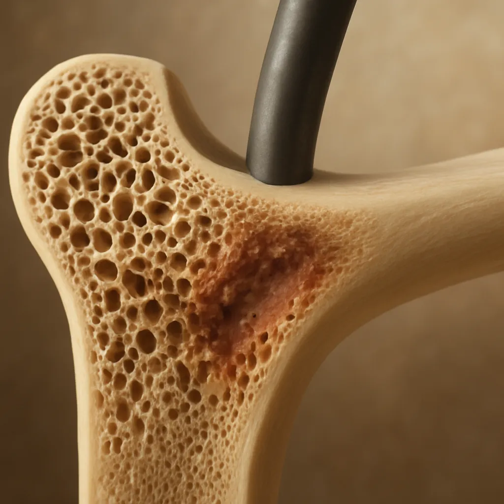
The organic matrix primarily consists of type I collagen, proteoglycans, and non-collagenous proteins, providing tensile strength and flexibility. Mineralization, predominantly by hydroxyapatite crystals, imparts compressive resistance. Mechanical loading alters matrix properties by inducing microcracks, which trigger targeted remodeling. This process maintains the integrity of the bone’s microarchitecture, preventing the accumulation of fatigue damage and preserving overall mechanical competence.
Mechanotransduction Mechanisms
Mechanotransduction refers to how bone cells detect and convert mechanical signals into biochemical responses. Strain induced by physical load activates a cascade of events that ultimately influence gene expression, cell proliferation, and matrix synthesis. The magnitude, rate, and frequency of mechanical input determine whether an anabolic response (bone formation) or a catabolic phase (resorption) prevails.
Cellular Sensors of Mechanical Load
- Primary cilia: Antennae-like structures on osteocytes that bend under fluid shear, triggering intracellular signaling.
- Ion channels: Stretch-activated channels such as Piezo1 allow Ca2+ influx in response to membrane deformation.
- Integrins: Transmembrane receptors connecting the cytoskeleton to the extracellular matrix, transmitting mechanical forces directly into biochemical cues.
- Focal adhesions: Complexes of proteins like talin and vinculin that anchor cells and relay mechanical stress to the actin cytoskeleton.
Signal Transduction Pathways
Once mechanical forces are detected, several intracellular pathways orchestrate the remodeling response:
- Wnt/β-catenin signaling: Enhances osteoblast differentiation and activity, promoting bone formation.
- MAPK and ERK cascades: Regulate proliferation and survival of bone cells.
- PI3K/Akt pathway: Mediates anti-apoptotic signals in osteocytes under mechanical strain.
- RhoA/ROCK signaling: Controls cytoskeletal tension and cell shape adjustments necessary for mechanosensation.
Together, these pathways integrate mechanical cues with hormonal and nutritional factors to adapt bone architecture appropriately.
Clinical Implications and Therapeutic Approaches
Insights into bone mechanobiology have profound implications for managing skeletal disorders, enhancing fracture healing, and optimizing athletic performance. Strategies that harness mechanical stimuli can complement pharmacological interventions to maximize bone strength and reduce fracture risk.
Orthopedic Interventions
Devices such as fixators and intramedullary nails apply controlled mechanical stimuli at fracture sites, stimulating callus formation and accelerating healing. Similarly, spinal fusion procedures rely on mechanical stability and local loading conditions to encourage bone growth across vertebral segments. Understanding the relationship between implant stiffness, stress shielding, and bone resorption guides the design of next-generation biomaterials that mimic the elastic properties of native bone.
Exercise and Physical Therapy
Weight-bearing and resistance exercises are key to increasing peak bone mass during growth and maintaining skeletal health in adulthood. Mechanical loading protocols are tailored to maximize osteogenic potential while minimizing injury risk. Physical therapists often prescribe:
- High-impact activities like jumping and plyometrics for young adults.
- Resistance band and weight-based training for older individuals to counteract age-related bone loss.
- Balance and proprioception exercises to reduce fall risk and subsequent fractures.
Emerging research on vibration therapy and whole-body vibration devices explores alternative methods to deliver mechanical stimuli to bone with low energy input.
Pharmacological Agents
Drugs targeting the remodeling process can amplify or moderate the effects of mechanical stress. Examples include:
- Bisphosphonates: Inhibit osteoclast-mediated resorption, effectively shifting the balance toward bone retention.
- Denosumab: A monoclonal antibody against RANKL that reduces osteoclast formation and activity.
- Teriparatide: An intermittent parathyroid hormone analog that preferentially stimulates mesenchymal stem cells to differentiate into osteoblasts, enhancing bone formation.
- Sclerostin inhibitors: Block the Wnt pathway antagonist sclerostin, promoting anabolic remodeling in response to load.
Combining pharmacotherapy with targeted exercise regimens yields synergistic benefits, enabling patients to achieve superior outcomes in bone density and structural quality.
Future Directions
Advances in tissue engineering and biomechanics promise novel solutions for skeletal repair. Bioactive scaffolds loaded with growth factors mimic the mechanical microenvironment of bone, guiding cell behavior through Wolff’s law. Computational models integrating finite element analysis predict strain distributions in patient-specific geometries, informing surgical planning and rehabilitation strategies. Continued exploration of mechanotransduction molecules may reveal new drug targets to modulate the remodeling process precisely.