




Bone metabolism is intricately regulated by the interplay between hormonal signals, cellular activity, and mineral homeostasis. In hyperparathyroidism, excessive secretion of parathyroid hormone (PTH) disrupts this delicate equilibrium, leading to alterations in bone remodeling dynamics and skeletal integrity. Understanding the mechanisms by which elevated PTH promotes bone resorption, the resulting clinical sequelae, and the optimal strategies to preserve bone strength is essential for effective patient management.
Pathophysiology of Hyperparathyroidism and Bone Remodeling
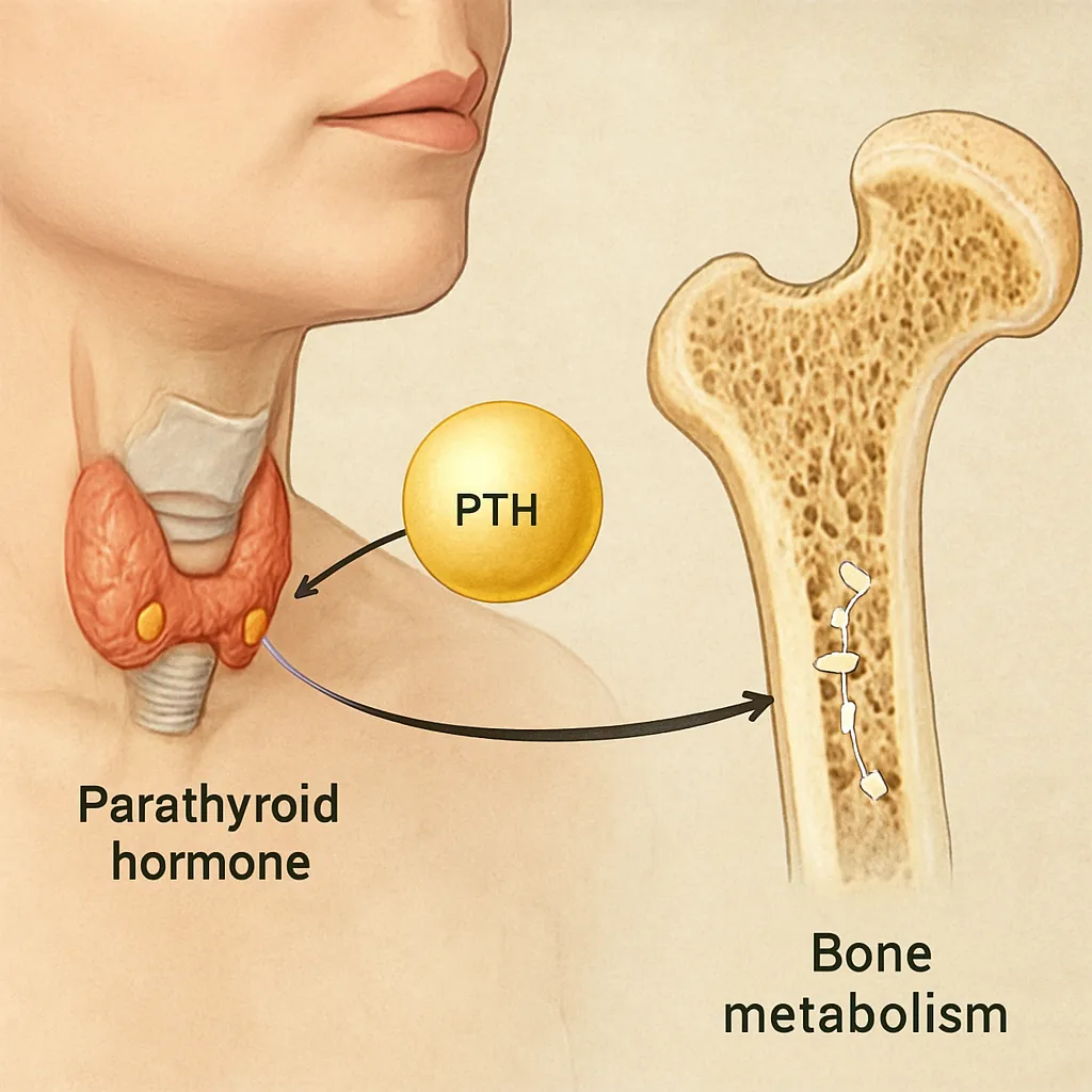
Parathyroid hormone, secreted by the parathyroid glands, is a principal regulator of calcium and phosphate balance. Under physiological conditions, PTH maintains serum calcium within a narrow range by stimulating renal reabsorption, activating vitamin D, and modulating bone turnover. In primary hyperparathyroidism, an autonomous adenoma or hyperplasia results in persistently elevated PTH levels. Secondary hyperparathyroidism arises from chronic hypocalcemia (commonly in chronic kidney disease), and tertiary hyperparathyroidism represents the transition to autonomous PTH secretion after prolonged secondary stimulation.
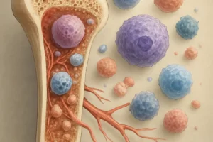
- Increased PTH binds to receptors on osteoblasts, upregulating RANKL (receptor activator of nuclear factor κB ligand).
- RANKL interacts with RANK on osteoclast precursors, enhancing their differentiation into mature osteoclasts.
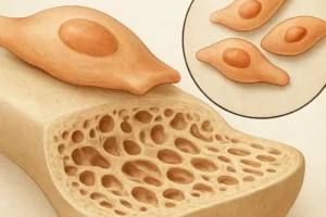
- Activated osteoclasts accelerate bone matrix degradation, leading to cortical thinning and trabecular perforation.
- Imbalances in osteoprotegerin (OPG), a decoy receptor for RANKL, further potentiate resorptive activity.
The net effect is a high-turnover state characterized by increased bone remodeling units. Markers such as serum alkaline phosphatase and urinary hydroxyproline rise in tandem with osteoclastic activity. Over time, chronic PTH exposure alters bone microarchitecture, reducing mechanical competence and predisposing to fragility fractures.
Clinical Manifestations and Skeletal Complications
Patients with hyperparathyroidism may present with a spectrum of symptoms ranging from subtle bone discomfort to overt skeletal deformities. The classic mnemonic “stones, bones, groans, and psychiatric overtones” encapsulates the multisystem involvement:
- Bone pain and tenderness, often localized to the long bones and ribs.
- Subperiosteal bone resorption, visible on radiographs as cortical thinning along the radial aspects of the middle phalanges.
- Brown tumors, focal lytic lesions resulting from osteoclastic overactivity and hemorrhagic infiltration.
- Pathological fractures at sites of high stress, including the vertebrae and femoral neck.
- Nephrolithiasis due to hypercalciuria, exacerbating renal parenchymal damage.
- Generalized muscle weakness, fatigue, and neuropsychiatric disturbances.
Advanced bone disease may manifest as distortions in vertebral shape, loss of height, and increased kyphosis. In secondary hyperparathyroidism, skeletal changes frequently involve the skull (“salt-and-pepper” appearance) and long bones (looser zones). Clinicians should remain vigilant for subtle signs on imaging studies to initiate timely intervention.
Diagnostic Modalities and Biomarkers
Accurate diagnosis hinges on correlating biochemical abnormalities with imaging findings. Laboratory evaluation typically includes:
- Serum calcium (total and ionized) – persistent elevation suggests primary hyperparathyroidism.
- Serum phosphate – often low in primary hyperparathyroidism and high in secondary forms.
- Parathyroid hormone levels – inappropriately elevated or normal despite hypercalcemia.
- 25-hydroxyvitamin D and 1,25-dihydroxyvitamin D – to identify concomitant deficiencies.
- Bone turnover markers (e.g., P1NP, CTX) – reflect the dynamic rate of bone formation and resorption.
Imaging plays a complementary role. Dual-energy X-ray absorptiometry (DEXA) quantifies bone mineral density at the lumbar spine, hip, and forearm, revealing the extent of osteopenia or osteoporosis. High-resolution peripheral quantitative computed tomography (HR-pQCT) can assess microarchitectural integrity, while dual-phase sestamibi scanning localizes hyperfunctioning parathyroid tissue. Ultrasonography, though operator-dependent, offers a noninvasive method to visualize enlarged glands. In select cases, bone biopsy may be warranted to characterize histological patterns of bone turnover.
Therapeutic Strategies for Bone Preservation
Management goals include normalization of serum calcium, mitigation of bone loss, and prevention of fractures. Treatment modalities encompass both pharmacological and surgical approaches:
Medical Management
- Calcimimetics (e.g., cinacalcet) enhance the sensitivity of calcium-sensing receptors on parathyroid cells, reducing PTH secretion and serum calcium.
- Bisphosphonates inhibit osteoclast-mediated resorption, improving bone density and lowering fracture risk.
- Selective estrogen receptor modulators (SERMs) may benefit postmenopausal women by promoting favorable bone remodeling.
- In secondary hyperparathyroidism, phosphate binders and active vitamin D analogs (e.g., calcitriol) help correct mineral disturbances.
- Dietary modifications: adequate calcium intake (1,000–1,200 mg/day) and avoidance of excessive dietary phosphorus.
Surgical Intervention
- Parathyroidectomy is the definitive therapy for symptomatic primary hyperparathyroidism or asymptomatic patients meeting criteria (e.g., serum calcium >1 mg/dL above normal, reduced bone density, nephrolithiasis).
- Minimally invasive approaches target a single adenoma, while subtotal or total parathyroidectomy with autotransplantation is reserved for hyperplasia.
- Postoperative monitoring focuses on calcium levels to detect hungry bone syndrome, characterized by sudden bone remineralization and hypocalcemia.
Long-term follow-up involves periodic assessment of bone density, renal function, and serum biomarkers. Multidisciplinary coordination with endocrinologists, nephrologists, and orthopedic specialists ensures a comprehensive strategy to preserve skeletal health in patients with hyperparathyroidism.