




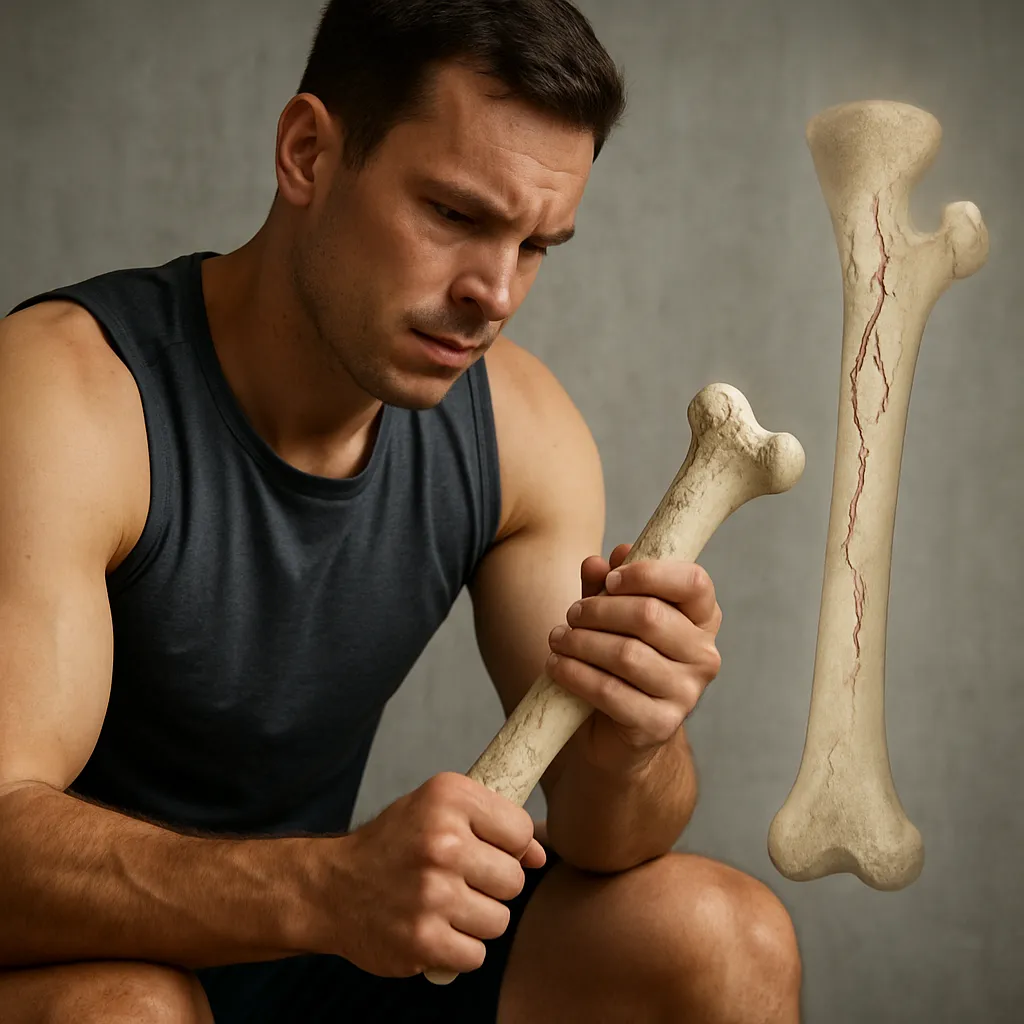
Bone fatigue in athletes represents a delicate balance between dynamic mechanical loads and the body’s intrinsic capacity for repair. As training intensity escalates, repetitive stresses can outpace the natural remodeling process, leading to the accumulation of microcracks and, ultimately, structural weakening. This phenomenon not only threatens performance but also raises the risk of long-term musculoskeletal complications. Understanding the interplay of physiological, biomechanical, and nutritional factors is essential for optimizing training regimens and safeguarding athlete health.
Physiology of Bone Fatigue
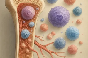
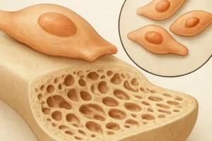
Bone tissue is constantly subjected to loads that trigger an adaptive response. Osteoblasts and osteoclasts coordinate the remodeling cycle, resorbing damaged matrix and depositing new mineralized tissue. However, when microdamage accumulates faster than repair can occur, the result is compromised integrity. Central to this process are osteocytes, the mechanosensing cells embedded within the bone matrix. They detect strain and signal for localized remodeling. With athletic training, cyclic loading promotes beneficial adaptation, but excessive repetition leads to fatigue cracks.
Microdamage Accumulation
Microdamage appears as tiny fissures within the collagen-mineral composite. Initially, the bone tolerates these defects, but if the defect density surpasses a critical threshold, it becomes a breeding ground for larger cracks. Researchers quantify this process using histological analysis, revealing that high-impact sports accelerate microcrack propagation. This underscores the importance of monitoring training volumes and incorporating rest intervals to allow for complete bone repair.
Bone Remodeling Cycle
The remodeling cycle involves distinct phases:
- Activation: Osteoclasts are recruited to sites of damage.
- Resorption: Damaged bone is broken down, releasing minerals.
- Reversal: Mononuclear cells prepare the surface for new matrix deposition.
- Formation: Osteoblasts synthesize new collagen and mineralize the matrix.
The duration of this cycle varies by skeletal site and individual factors such as hormonal status and nutrient availability.
Contributing Factors in Athletes
Athletes encounter unique demands that predispose them to bone fatigue. The magnitude of repetitive loads, training frequency, and movement patterns significantly influence stress distribution.
Biomechanical Stressors
Running, jumping, and cutting maneuvers impose distinct stress profiles on weight-bearing bones. Adequate muscle strength and joint alignment help dissipate forces. However, biomechanical inefficiencies—such as excessive pronation or limb length discrepancies—can concentrate stress on focal bone areas. Continuous monitoring of gait patterns and implementing corrective exercises are crucial for reducing localized overload.
Nutritional and Hormonal Influences
Optimal bone health depends on a balanced supply of calcium, vitamin D, and protein. Insufficient dietary intake impairs mineralization and weakens structural resilience. Additionally, endocrine factors such as estrogen and testosterone play pivotal roles in regulating remodeling rates. Female athletes with menstrual irregularities may experience reduced bone density, heightening susceptibility to stress fractures.
Training Load and Recovery
Progressive overload drives adaptation, but inadequate recovery fosters microdamage accumulation. Training programs should integrate periodization, alternating high-intensity sessions with active rest. Sleep and rest days facilitate cellular repair mechanisms, allowing osteoblasts to complete the formation phase without interruption.
Diagnostic and Imaging Tools
Early detection of bone fatigue is vital to prevent progression to frank fractures. Clinicians employ a combination of clinical assessment and advanced imaging modalities.
Clinical Examination
History-taking focuses on pain onset, training changes, and anatomical location. Physical examination may reveal point tenderness, swelling, or gait alterations. Functional tests assess load tolerance and identify compensatory movement patterns.
Imaging Modalities
- X-ray: Useful for advanced stress injuries but insensitive to early microdamage.
- Magnetic Resonance Imaging (MRI): Highly sensitive for detecting bone marrow edema, an early sign of stress reaction.
- Bone Scintigraphy: Highlights areas of increased metabolic activity, indicating active remodeling.
- Computed Tomography (CT): Provides detailed cortical assessment but involves higher radiation exposure.
Emerging techniques like quantitative ultrasound and finite element analysis offer noninvasive insight into bone quality and mechanical competence.
Biochemical Markers
Laboratory assays measuring collagen breakdown products (e.g., N-terminal telopeptide) and osteoblastic activity markers (e.g., bone-specific alkaline phosphatase) provide indirect evidence of remodeling dynamics. These biomarkers help tailor training intensity by indicating whether the bone turnover rate is balanced or skewed toward resorption.
Strategies for Prevention and Management
Proactive measures can mitigate the risk of bone fatigue and facilitate faster recovery when issues arise. A multifaceted approach addresses mechanical, nutritional, and physiological factors.
Optimizing Training Programs
Implementing a periodized plan ensures gradual load progression and incorporates de-loading phases. Cross-training reduces repetitive stress by diversifying movement patterns. Strength and conditioning protocols targeting core stability and lower limb alignment improve force distribution and reduce focal overload.
Nutrition and Supplementation
A nutrient-rich diet supports mineralization and collagen synthesis. Key recommendations include:
- Adequate calcium intake (1,000–1,300 mg/day).
- Vitamin D optimization (800–2,000 IU/day), monitored via serum 25(OH)D levels.
- Sufficient high-quality protein (1.2–2.0 g/kg body weight daily).
- Consideration of collagen or gelatin supplements to enhance matrix quality.
Proper hydration also maintains the viscoelastic properties of bone and surrounding soft tissues.
Recovery and Rehabilitation
When early signs of bone fatigue appear, immediate load reduction is imperative. Rehabilitation focuses on pain-free mobility exercises and gradual reintroduction of weight-bearing activities. Aquatic therapy and low-impact cross-training maintain cardiovascular fitness without compromising bone integrity.
Stress Fracture Susceptibility
Certain skeletal sites, such as the tibial shaft, metatarsals, and femoral neck, demonstrate higher vulnerability. Classification systems grade stress injuries from periosteal reactions to complete fractures. Treatment duration correlates with injury grade, ranging from weeks of immobilization to surgical intervention for displaced or high-risk lesions.
Monitoring and Return-to-Play Criteria
Objective measures guide the safe resumption of sport:
- Absence of pain during functional tests.
- Normalization of imaging findings or biomarkers.
- Completion of a structured return-to-play progression without symptoms.
Regular follow-up ensures that underlying risk factors are addressed before full competitive load is restored.