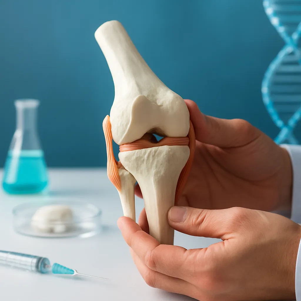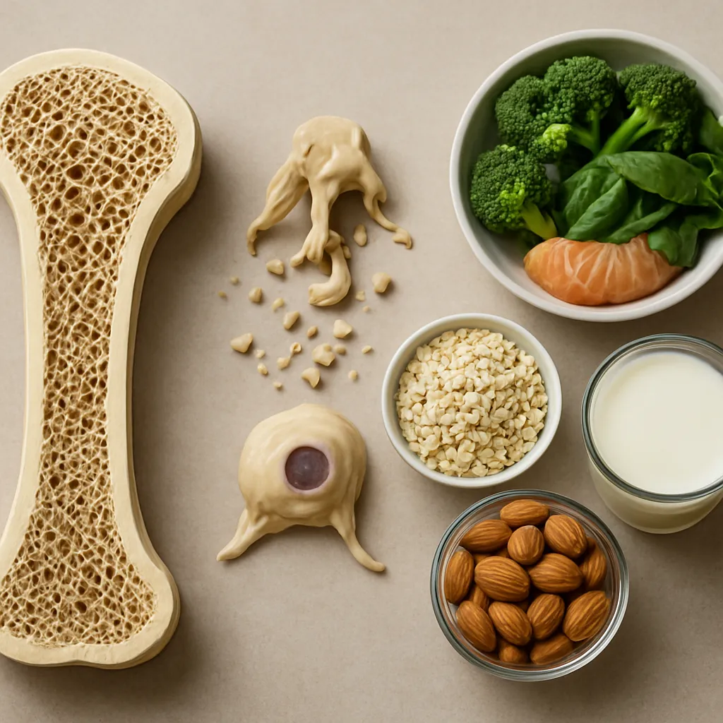



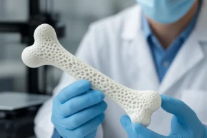

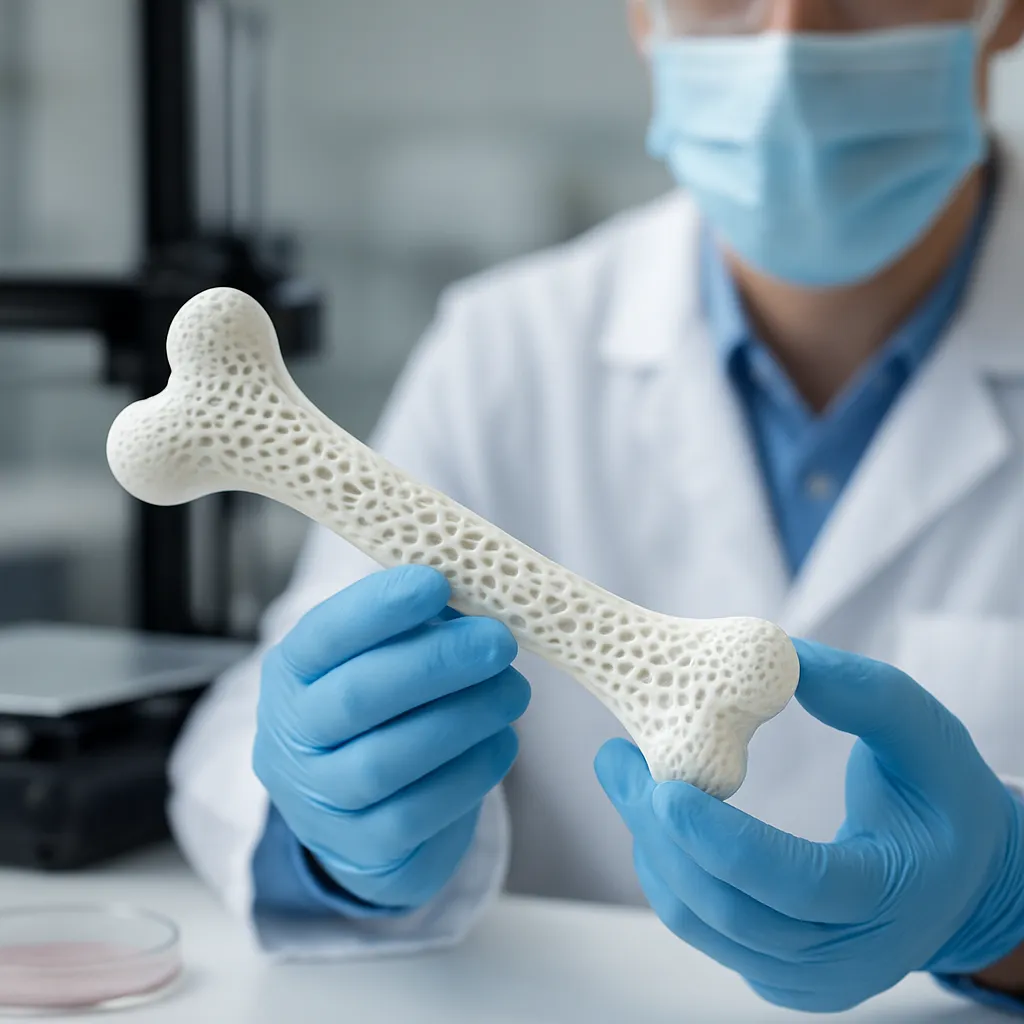
The rapid evolution of tissue engineering is setting the stage for a paradigm shift in orthopedic medicine. By harnessing the power of bioprinting technology, researchers are developing patient-specific implants that promise to restore function and accelerate healing. This article delves into the breakthroughs, methodologies, and hurdles encountered on the path toward fully functional, 3D-printed bone replacements.
Advancements in Biomaterials for 3D Bioprinting
Central to any successful bone substitute is the composition of the bioinks and scaffold materials. Over the past decade, scientists have refined a slew of biomaterials to mimic the native environment of bone tissue. By adjusting the ratio of polymers, ceramics, and growth factors, these composite formulations can facilitate osteogenesis and support cellular adhesion.
Composite Scaffolds and Their Role
- Hydroxyapatite and tricalcium phosphate: Minerals that closely resemble natural bone matrix.
- Biodegradable polymers such as polycaprolactone (PCL) and polylactic acid (PLA): Provide structural support while gradually degrading as new tissue forms.
- Gelatin methacrylate (GelMA) and alginate blends: Offer a hydrated environment that fosters nutrient exchange and cell proliferation.
Innovations in composite scaffolds are enabling constructs with finely tuned mechanical strength, essential for load-bearing applications. By layering ceramics within a polymer framework, engineers can create implants that bridge the gap between rigidity and flexibility, reducing the risk of stress shielding.
Growth Factors and Cellular Components
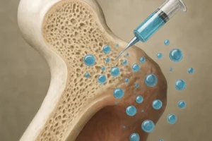
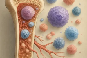
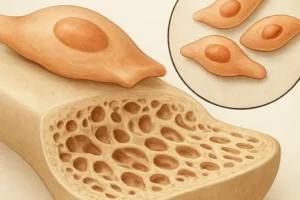
The addition of bioactive molecules such as bone morphogenetic proteins (BMPs) and vascular endothelial growth factor (VEGF) has been transformative. These signaling proteins guide stem cell differentiation and enhance vascularization, ensuring that newly forming bone receives adequate blood supply. Moreover, seeding scaffolds with mesenchymal stem cells (MSCs) or osteoblast precursors further promotes tissue regeneration, steering the construct toward functional integration with the host.
Techniques and Technologies Shaping Bone Fabrication
Multiple 3D printing modalities have been adapted to create intricate bone constructs. Each technique offers unique advantages in resolution, speed, and material compatibility, expanding the toolbox available to tissue engineers.
Extrusion-Based Bioprinting
By dispensing continuous filaments of material, extrusion systems excel at producing large, porous structures. Temperature-controlled nozzles enable precise deposition of thermoplastic polymers, while pneumatic or screw-driven mechanisms handle viscous bioinks laden with cells and growth factors.
Inkjet and Laser-Assisted Approaches
Inkjet bioprinters propel droplets of cell-laden solutions onto a substrate with high accuracy, making them ideal for patterning delicate cellular arrays. Laser-assisted bioprinting, on the other hand, uses focused energy to transfer biomaterial from a donor ribbon to a collector surface, offering exceptional resolution without nozzle clogging.
Digital Light Processing (DLP) and Stereolithography
Photo-curable resins form the basis of DLP-based systems, which can solidify entire layers with a single exposure to patterned light. This voxel-by-voxel approach yields smooth surfaces and complex internal channels, critical for designing implants that mimic the hierarchical organization of bone.
Clinical Applications and Challenges
While preclinical studies have demonstrated promising outcomes, translating 3D-printed bone replacements into human patients remains a multifaceted endeavor. Researchers must navigate biological, regulatory, and logistical hurdles before widespread adoption can occur.
Preclinical Validation and Animal Models
Rodent and large-animal studies have provided valuable insights into the in vivo performance of bioprinted constructs. Successful graft integration, bone remodeling, and restoration of mechanical function have been observed in rabbit femoral defects and sheep spinal fusion models. Yet, scaling up these findings to address the variability found in human anatomy and physiology is a substantial task.
Immune Response and Biocompatibility
The host immune reaction to implanted materials can either support healing or lead to chronic inflammation and implant failure. By selecting regenerative polymers that degrade into non-toxic by-products, researchers aim to minimize adverse responses. Coating scaffolds with immunomodulatory agents or employing autologous cells also helps in mitigating rejection risks.
Regulatory and Manufacturing Hurdles
Achieving clinical translation requires adherence to stringent quality standards. Good Manufacturing Practice (GMP) guidelines demand impeccable traceability of raw materials, reproducible printing protocols, and rigorous sterility controls. Additionally, establishing cost-effective workflows for custom-designed implants remains a significant barrier, particularly in healthcare systems with limited resources.
Future Directions in Bone Bioprinting
Looking ahead, the convergence of digital imaging, artificial intelligence, and bioreactor technologies is poised to accelerate innovation in bone replacement therapies.
Personalized Implants via Imaging Integration
Advanced medical imaging modalities such as high-resolution CT and MRI enable precise mapping of bone defects. By integrating these images with computer-aided design (CAD) software, engineers can generate customization plans that account for patient-specific geometry and load-bearing requirements. Automated segmentation and optimization algorithms further refine implant designs, ensuring optimal fit and functionality.
Machine Learning for Bioink Formulation
Artificial intelligence platforms are being trained to predict the printability and biological performance of novel bioink recipes. By analyzing large datasets of material properties and cellular outcomes, machine learning models can suggest formulations that balance viscosity, crosslinking kinetics, and nutrient diffusion, reducing the trial-and-error cycle in laboratory settings.
Bioreactors and Dynamic Conditioning
To mimic the mechanical and biochemical cues present in the body, 3D-printed constructs can be matured within bioreactors that apply cyclic loading, shear stress, and controlled perfusion. These dynamic environments stimulate cellular remodeling and extracellular matrix deposition, priming implants for successful integration upon transplantation.
As the field continues to evolve, collaboration among materials scientists, biologists, engineers, and clinicians will be paramount. The quest for fully functional, patient-tailored bone replacements is not solely a technological challenge but a multidisciplinary journey that holds the promise of transforming the landscape of orthopedic care.