Bone graft integration and vascularization are fundamental components of successful reconstructive procedures in orthopedics and maxillofacial surgery. Achieving stable incorporation of grafts into host bone depends on multiple biological and mechanical factors that govern graft survival, new bone formation, and long-term functional outcomes. Understanding these principles supports the development of advanced biomaterials and therapeutic strategies to optimize healing and patient recovery.
Bone Graft Materials and Biological Principles
Autografts and Allografts
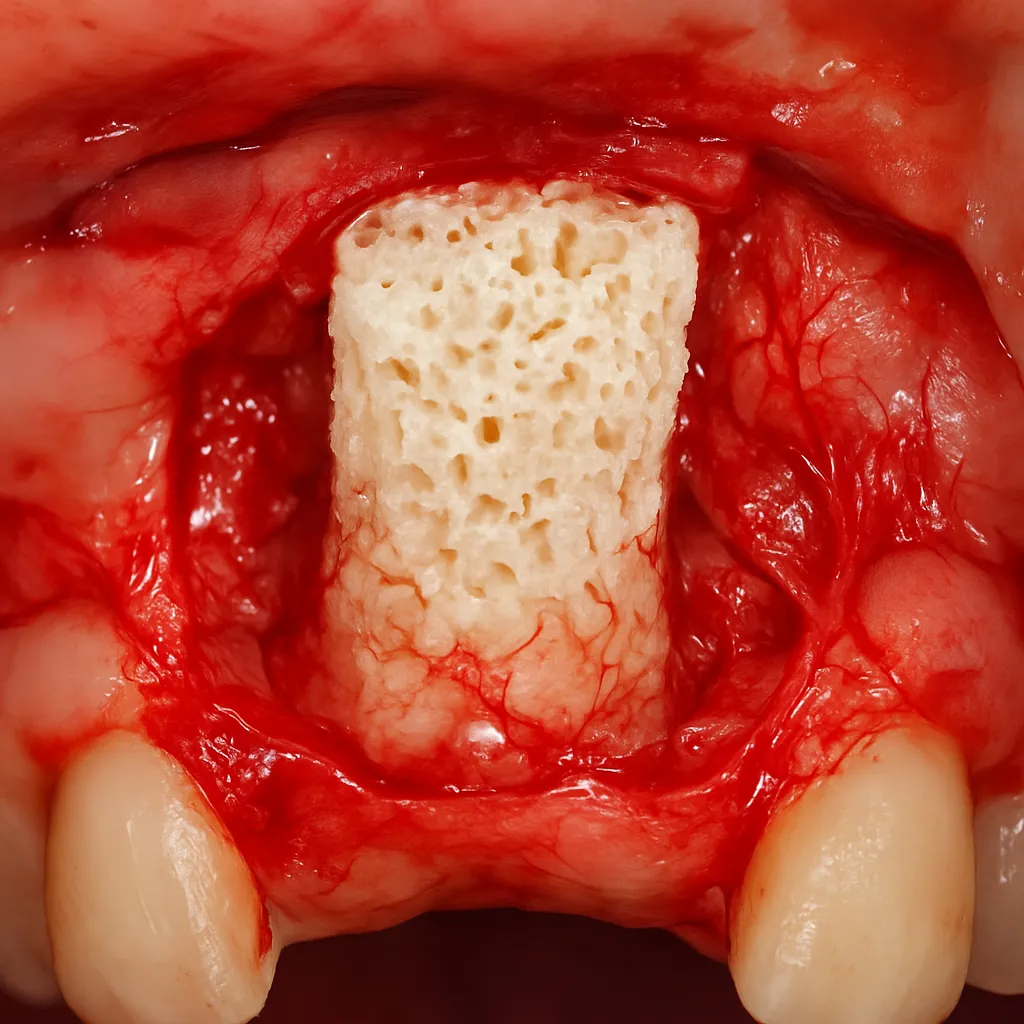
Autologous bone grafts, harvested from the patient’s own body, remain the clinical gold standard because of their inherent osteogenic, osteoinductive, and osteoconductive properties. The presence of living osteoblasts and progenitor cells fosters rapid osseointegration without eliciting an immune reaction. Allografts, sourced from human donors, offer abundant supply and eliminate donor-site morbidity. However, processing methods such as freeze-drying or gamma irradiation can compromise biocompatibility and reduce the biological activity of native proteins within the graft.
Synthetic Scaffolds and Bioceramics
Synthetic graft substitutes, including hydroxyapatite, β-tricalcium phosphate, and bioactive glass, serve as scaffold materials to guide new bone deposition. Their porosity and interconnectivity are engineered to mimic the natural extracellular matrix, facilitating cell migration and nutrient diffusion. Advances in polymer-ceramic composites have improved mechanical strength and controlled degradation kinetics. Yet, achieving optimal vascularization within these constructs remains a key challenge to prevent central necrosis and ensure uniform bone remodeling.
Mechanisms of Vascular Infiltration and Osteogenesis
Role of Angiogenic Factors
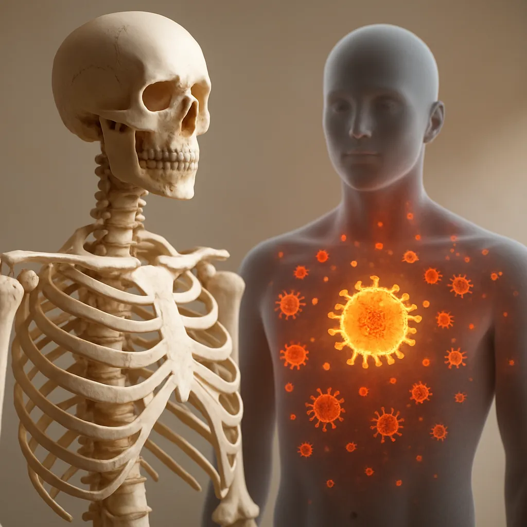
Successful graft integration relies on coordinated angiogenesis to supply oxygen, nutrients, and growth factors. Vascular endothelial growth factor (VEGF), basic fibroblast growth factor (bFGF), and platelet-derived growth factor (PDGF) act synergistically to promote endothelial cell proliferation and migration. Local delivery of these cytokines, either via impregnated scaffolds or microsphere systems, has demonstrated accelerated capillary ingrowth and enhanced bone formation in preclinical models.
Cellular Contributors
Mesenchymal stem cells (MSCs) and endothelial progenitor cells (EPCs) play central roles in orchestrating the interplay between new vessel formation and bone tissue regeneration. MSCs differentiate into osteoblasts under osteoinductive stimuli, while EPCs line nascent capillaries, creating a microenvironment that supports continued remodeling. Co-culture systems and three-dimensional bioreactors enable researchers to study the dynamic crosstalk between these cell populations and biomaterial matrices in vitro, guiding design improvements for in vivo applications.
Clinical Strategies to Enhance Integration
Growth Factor Delivery Systems
Innovative delivery platforms aim to provide sustained release of bioactive molecules at the graft site. Thermoresponsive hydrogels, nanoparticle carriers, and layer-by-layer coatings on scaffold surfaces offer temporal control over factor presentation. By finely tuning release profiles, clinicians can synchronize angiogenic peaks with osteoblastic activity, maximizing vascularization while minimizing potential side effects of supraphysiological doses.
Advanced Tissue Engineering Approaches
Three-dimensional bioprinting enables fabrication of patient-specific grafts that incorporate channels for perfusion and gradients of mechanical properties. Customizable architectures facilitate directional vessel in-growth and load-bearing function. Moreover, gene-activated matrices, which deliver plasmids encoding for VEGF or BMP-2, harness the host’s own cells to produce therapeutic proteins in situ. Such approaches hold promise for overcoming limitations of traditional grafts and reducing reliance on exogenous growth factors.
Future Directions and Research Frontiers
Emerging research focuses on integrating smart materials that respond to environmental cues, such as pH and enzymatic activity, to modulate degradation and release of encapsulated agents. Nanostructured coatings that mimic natural bone topography can further guide cell behavior. Innovations in imaging, including intravital microscopy, allow real-time monitoring of graft vascularization and cellular dynamics. Interdisciplinary collaborations among material scientists, cell biologists, and surgeons are crucial to translate these discoveries into clinical practice, ultimately improving patient outcomes and expanding the therapeutic potential of bone grafting techniques.