Bone density recovery after a fracture is a complex, tightly regulated process that requires the coordinated action of various cellular players, biochemical signals, mechanical stimuli, and patient-specific factors. Understanding these interactions is critical for developing effective interventions that accelerate healing, prevent complications, and restore full function. This article explores the underlying physiology, identifies key influencers on recovery, and outlines evidence-based strategies to optimize bone regeneration.
Bone Healing Physiology
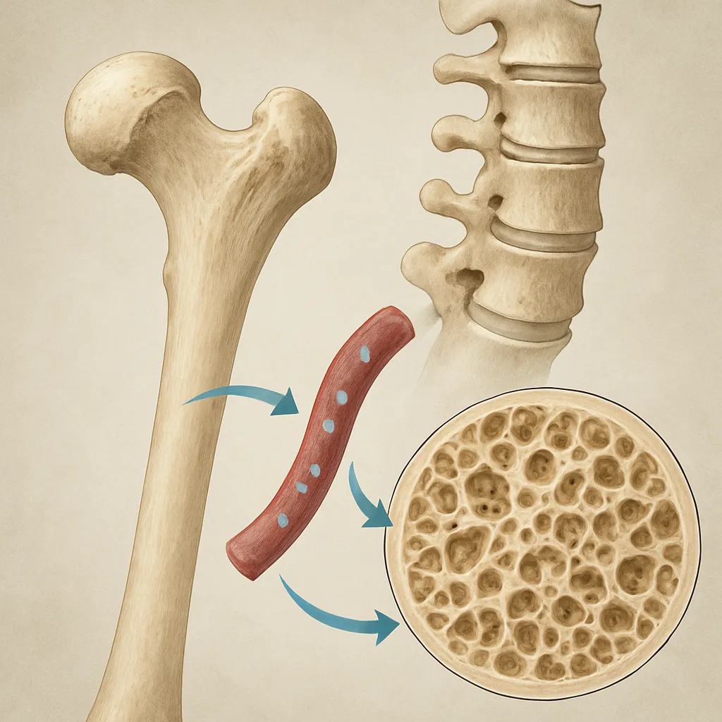
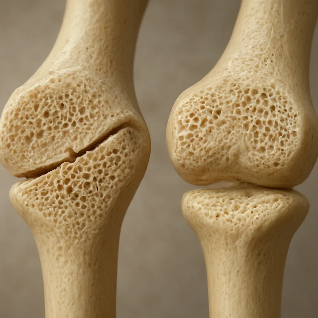
Bone healing unfolds in three partially overlapping phases: inflammation, repair, and remodeling. Immediately after a fracture, damaged vessels release cytokines and growth factors that attract immune cells and promote hematoma formation. In the repair phase, osteoblasts and chondrocytes drive callus formation, laying down a combination of cartilaginous and woven bone. Finally, during remodeling, osteoclasts resorb immature bone while osteoblasts deposit lamellar bone, restoring the original architecture and strength.
Stage 1: Inflammatory Response
- Vascular disruption leads to hematoma rich in platelets and fibrin
- Secretion of interleukins (IL-1, IL-6) and tumor necrosis factor-alpha (TNF-α)
- Recruitment of mesenchymal stem cells (MSCs) to the fracture site
Stage 2: Reparative Phase
Within days, MSCs differentiate into osteoblasts and chondrocytes, forming a soft callus that bridges the gap. Mineralization follows, transforming it into a hard callus. Local levels of bone morphogenetic proteins (BMPs) and transforming growth factor-beta (TGF-β) orchestrate matrix synthesis.
Stage 3: Remodeling Phase
Over months, the hard callus is remodeled via the coupled actions of osteoclasts and osteoblasts, influenced by mechanical loading and systemic hormones. Mechanotransduction pathways, mediated by the Wnt/β-catenin signaling and sclerostin inhibition, guide adaptation to stress, ensuring the bone regains pre-injury resilience.
Factors Influencing Bone Density Recovery
Not every fracture heals at the same rate or with equal quality. Patient age, comorbidities, lifestyle choices, and fracture characteristics all play pivotal roles. Optimizing these variables can significantly affect the pace and completeness of bone restoration.
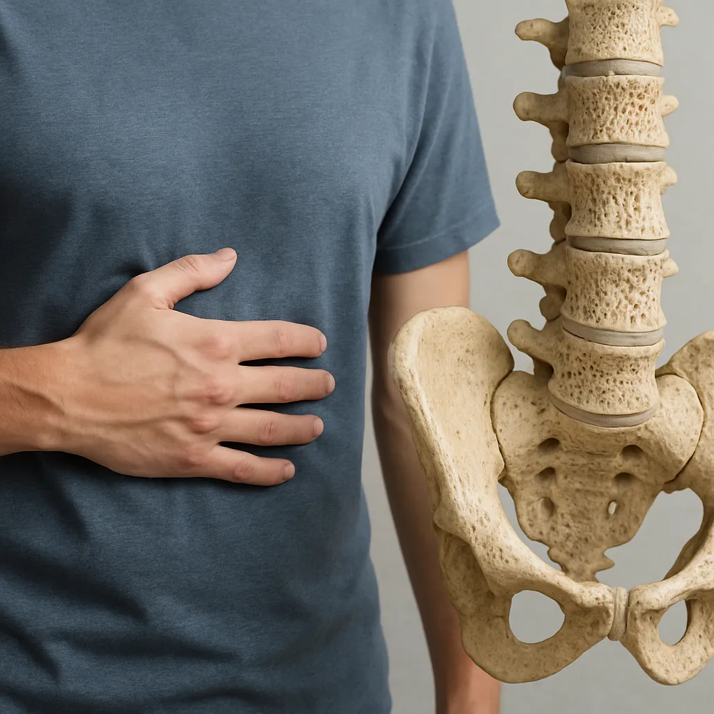
Age and Hormonal Status
With aging, there is a decline in MSC proliferative capacity and a shift in differentiation toward adipogenesis at the expense of osteogenesis. Postmenopausal estrogen deficiency exacerbates bone loss by increasing RANKL expression, favoring osteoclast activity. Consequently, elderly patients often exhibit slower callus formation and weaker remodeling.
Nutrition and Micronutrients
- Calcium: Essential for hydroxyapatite crystal formation.
- Vitamin D: Enhances intestinal calcium absorption and regulates mineral homeostasis.
- Protein: Supplies amino acids for collagen matrix synthesis.
- Trace elements (magnesium, zinc): Cofactors for enzymatic processes in bone metabolism.
Mechanical Loading and Rehabilitation
Controlled mechanical stimuli, through weight-bearing or low-intensity vibration, activate mechanotransduction pathways that upregulate osteogenic genes. Delayed or insufficient loading can lead to disuse osteopenia, while premature overload risks nonunion or malunion. A tailored rehabilitation program balances protection with progressive stress application.
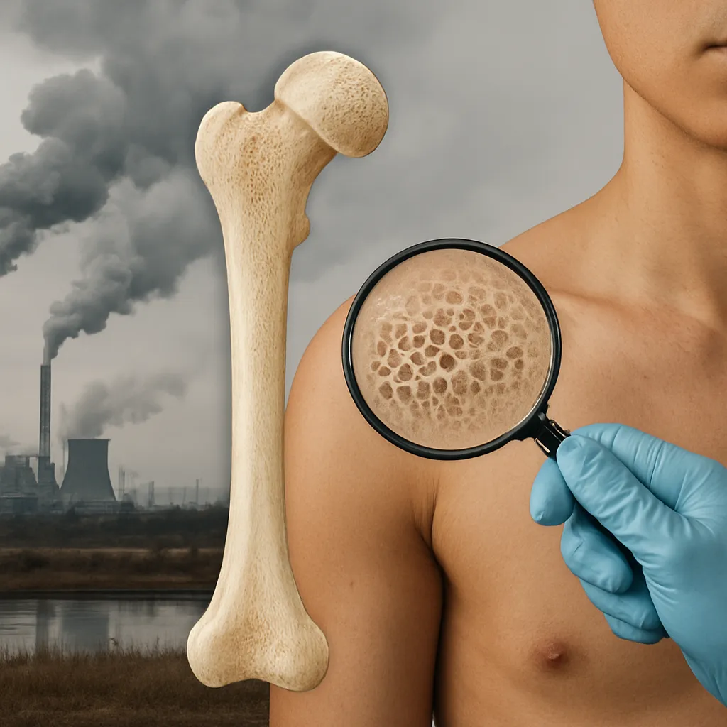
Systemic Conditions and Medications
Comorbidities such as diabetes, rheumatoid arthritis, and chronic kidney disease disrupt bone remodeling via altered cytokine profiles and impaired MSC function. Glucocorticoids, proton-pump inhibitors, and certain antiepileptic drugs can further diminish bone density and delay healing.
Advanced Strategies to Enhance Recovery
Modern medicine offers an expanding toolkit to support bone repair beyond standard immobilization. Strategies range from targeted pharmacologics to cutting-edge biologics and mechanical interventions.
Pharmacological Agents
- Bisphosphonates: Inhibit osteoclast-mediated resorption, preserving callus integrity. Timing is crucial; early administration may impair remodeling.
- Denosumab: A RANKL inhibitor that reduces bone turnover; used cautiously in fracture settings.
- Anabolic agents: Teriparatide (PTH analog) stimulates osteoblast activity and improves callus quality. Clinical trials show accelerated healing in osteoporotic fractures.
Biologics and Growth Factors
Recombinant BMP-2 and BMP-7 have FDA approval for select open fractures and spinal fusions. They enhance local MSC differentiation and matrix deposition. Platelet-rich plasma (PRP) and autologous stem cell concentrates are also under investigation, though protocols and outcomes remain variable.
Mechanical Stimulation Devices
- Low-intensity pulsed ultrasound (LIPUS): Thought to boost angiogenesis and osteogenesis; meta-analyses show modest reductions in healing time.
- Electrical bone growth stimulators: Deliver pulsed electromagnetic fields that may promote cell proliferation and matrix synthesis.
- Vibration platforms: Low-magnitude, high-frequency vibration supports trabecular bone maintenance in immobilized limbs.
Clinical Rehabilitation Protocols
Integrating physical therapy from an early postoperative stage fosters functional recovery and bone adaptation. Key elements include:
- Progressive weight-bearing: Starting with partial support and advancing to full load over weeks.
- Range-of-motion exercises: Prevent joint stiffness and encourage nutrient diffusion in peri-fracture tissues.
- Muscle strengthening: Particularly around the fracture site to decrease undue stress and improve stability.
- Proprioceptive training: Enhances neuromuscular control, reducing the risk of re-injury.
Close monitoring via serial imaging (X-ray, CT, or DEXA scans) allows clinicians to adjust the rehabilitation timeline based on callus maturity and callus density.
Emerging Research and Future Directions
Novel approaches, such as gene therapy targeting sclerostin suppression, show promise in preclinical models. Nanocomposite scaffolds impregnated with growth factors aim to deliver sustained, localized osteoinductive cues. Personalized medicine, informed by genetic profiling of bone turnover markers, could tailor interventions to individual healing phenotypes, maximizing outcomes and minimizing adverse effects.
As our understanding deepens, combining biomechanical insights with molecular innovations will forge more effective, patient-centered protocols for achieving rapid and robust bone density recovery after fracture.