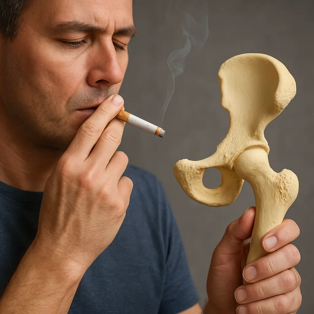
Smoking has long been recognized as a major risk factor for various chronic diseases, but its impact on the skeletal system is equally alarming. Beyond the well-documented harm to the lungs and cardiovascular system, tobacco use exerts deleterious effects on bone health, leading to decreased bone density, impaired healing, and heightened susceptibility to fractures. Fortunately, ceasing tobacco consumption triggers a cascade of beneficial physiological changes that can reverse or mitigate these negative outcomes. This article delves into the interplay between smoking and bone physiology, highlights the mechanisms by which quitting enhances skeletal integrity, reviews key clinical evidence, and offers practical strategies for optimizing bone health in former smokers.
Pathophysiological Mechanisms of Smoking-Induced Bone Damage
Tobacco smoke contains thousands of chemical constituents, including nicotine, carbon monoxide, and a myriad of free radicals. These agents undermine bone homeostasis through several interrelated pathways:
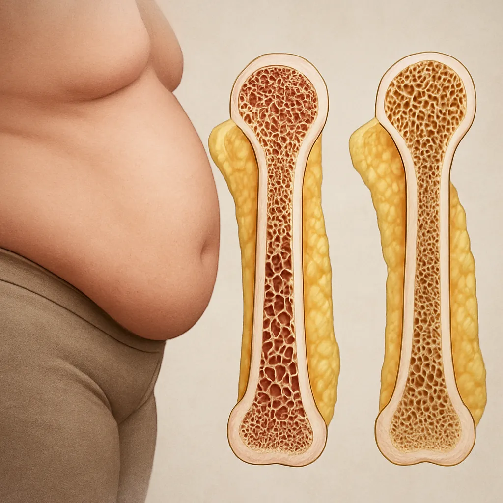
- Oxidative stress: Reactive oxygen species generated by smoke accelerate the apoptosis of osteoblasts, the cells responsible for new bone formation, while promoting osteoclast activity, which enhances bone resorption.
- Hormonal disruption: Smoking reduces levels of circulating estrogen in women and testosterone in men, both of which are crucial for maintaining bone mass and regulating remodeling cycles.
- Calcium absorption impairment: Nicotine alters gastrointestinal function and vitamin D metabolism, decreasing the efficiency of calcium uptake and thus limiting the mineral substrate needed for bone mineralization.
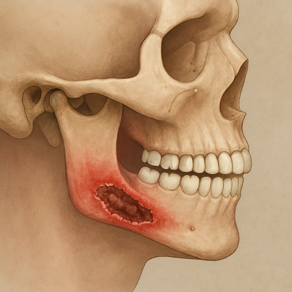
- Microvascular compromise: Carbon monoxide and other vasoconstrictive components diminish blood flow to bone tissue, slowing healing and reducing nutrient delivery.
Collectively, these factors drive a net bone loss state, elevating the risk of osteoporosis and fragility fractures, particularly in the hip, spine, and wrist regions.
Reversal of Bone Detriment Following Smoking Cessation
When an individual stops smoking, the body initiates repair processes that gradually restore bone health:
- Reduction of inflammatory markers: Levels of cytokines such as IL-6 and TNF-alpha decline, creating a more favorable environment for osteoblast function.
- Normalization of hormone levels: Estrogen and testosterone equilibrate, supporting the maintenance of existing bone mass and facilitating deposition of new matrix.
- Enhanced nutrient uptake: Improved vitamin D status and gastrointestinal integrity boost calcium absorption, supplying minerals necessary for bone mineral density gains.
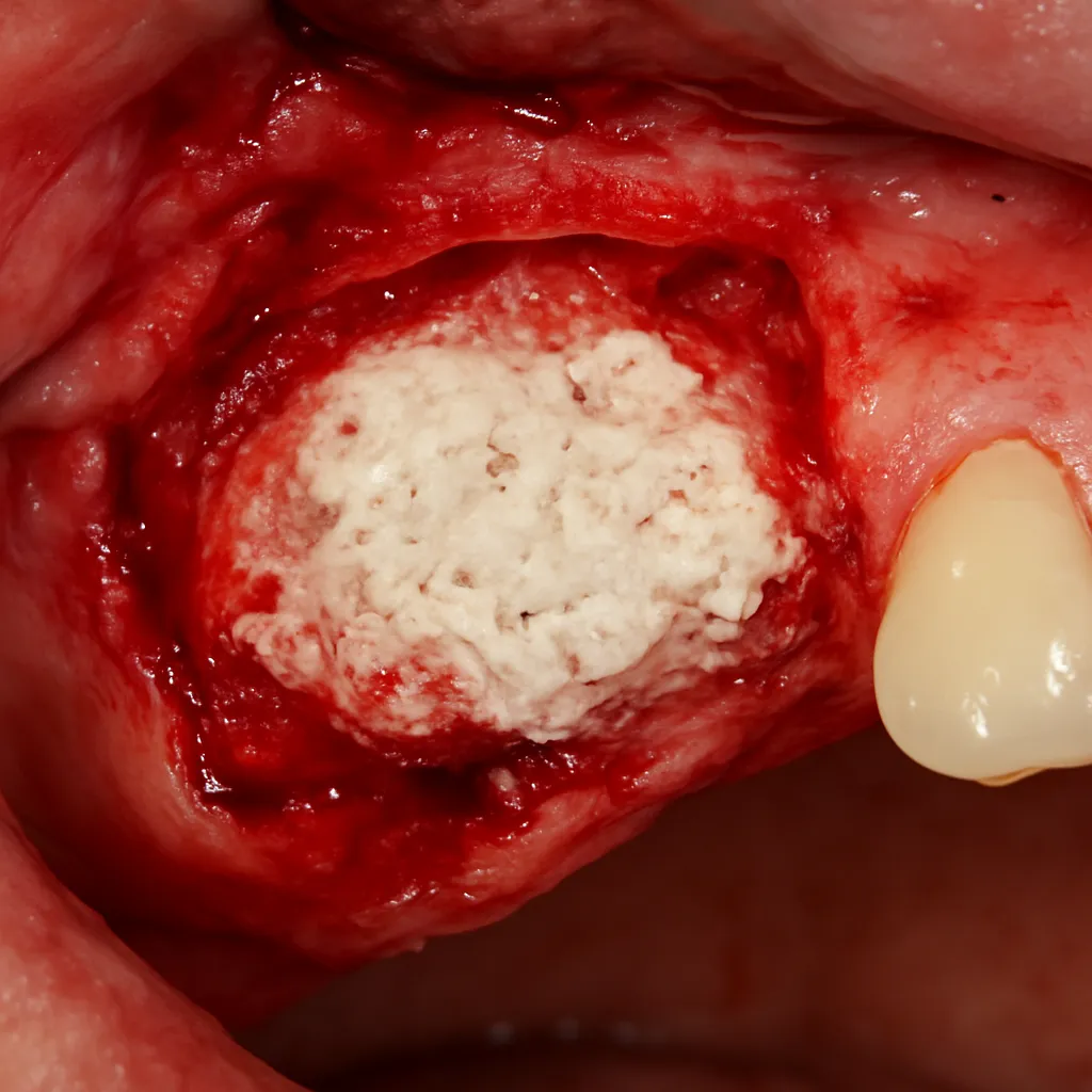
- Improved blood perfusion: Reversal of smoke-induced vasoconstriction enhances microcirculation in bone tissue, expediting repair and remodeling processes.
Studies show that within 6 to 12 months of quitting, markers of bone turnover begin to shift toward a more anabolic profile. Over longer durations, former smokers can achieve bone densities that approach those of never-smokers, significantly reducing fracture risk.
Clinical Evidence Supporting Bone Recovery
Numerous epidemiological and interventional trials have documented the skeletal benefits of smoking cessation:
Population-Based Cohort Studies
Large-scale cohorts reveal that former smokers experience a gradual decline in fracture incidence over time. In women over 50, the hazard ratio for hip fractures drops by approximately 20% after five years of cessation compared with continuous smokers. Similar trends appear in male cohorts, underscoring the universal advantage of quitting.
Bone Density Assessments
Dual-energy X-ray absorptiometry (DXA) studies demonstrate significant gains in lumbar spine and femoral neck bone mineral density (BMD) among individuals who quit smoking. In one prospective trial, participants who abstained for two years showed a mean BMD increase of 3–5% versus a continued-loss trajectory in persistent smokers.
Bone Turnover Biomarkers
Levels of osteocalcin (a marker of bone formation) and C-terminal telopeptide (CTX, a resorption marker) shift favorably after smoking cessation. This biochemical evidence aligns with histomorphometric findings of improved trabecular architecture in former smokers, indicating true structural gains beyond mere density measurements.
Strategies to Maximize Bone Health Post-Cessation
Cessation alone provides a robust foundation for skeletal recovery, but additional interventions can further enhance outcomes:
- Nutrition optimization: A diet rich in calcium (1,000–1,200 mg/day), vitamin D (800–1,000 IU/day), and protein supports matrix synthesis and mineralization.
- Weight-bearing exercise: Resistance training, walking, and impact activities stimulate osteoblast activity and improve bone geometry.
- Supplementation: In cases of documented deficiency, calcium and vitamin D supplements can correct insufficiencies and promote BMD gains.
- Pharmacotherapy: For individuals with established osteoporosis, medications such as bisphosphonates or denosumab may be indicated; smoking cessation enhances the efficacy of these agents.
- Lifestyle modifications: Limiting alcohol intake and ensuring adequate sleep further optimize the hormonal milieu and support bone remodeling.
Healthcare providers should include bone health assessments in smoking cessation programs, integrating risk stratification, DXA screening, and personalized intervention plans to safeguard skeletal wellness in this vulnerable population.
Future Directions in Research and Practice
While current evidence strongly supports the bone-protective effect of quitting smoking, several avenues warrant further exploration:
- Identification of genetic polymorphisms that influence the magnitude of bone recovery post-cessation.
- Long-term comparative trials evaluating different cessation aids (nicotine replacement, varenicline, bupropion) on bone turnover markers.
- Development of integrated care models that combine smoking cessation counseling with osteoporosis prevention strategies.
- Advanced imaging techniques, such as high-resolution peripheral quantitative computed tomography (HR-pQCT), to monitor microarchitectural repair in former smokers.
Advancing these research fronts will refine clinical guidelines and ensure that individuals who quit smoking reap the maximum possible benefit for their skeletal health.