





Bone density testing plays a pivotal role in identifying individuals at risk of developing osteoporosis and related complications. This article explores the science behind common screening methods, dispels widespread misconceptions, and provides evidence-based recommendations for clinicians and patients. By distinguishing myths from facts, healthcare professionals can optimize preventive strategies and improve patient outcomes.
Understanding the Principles of Bone Density Testing
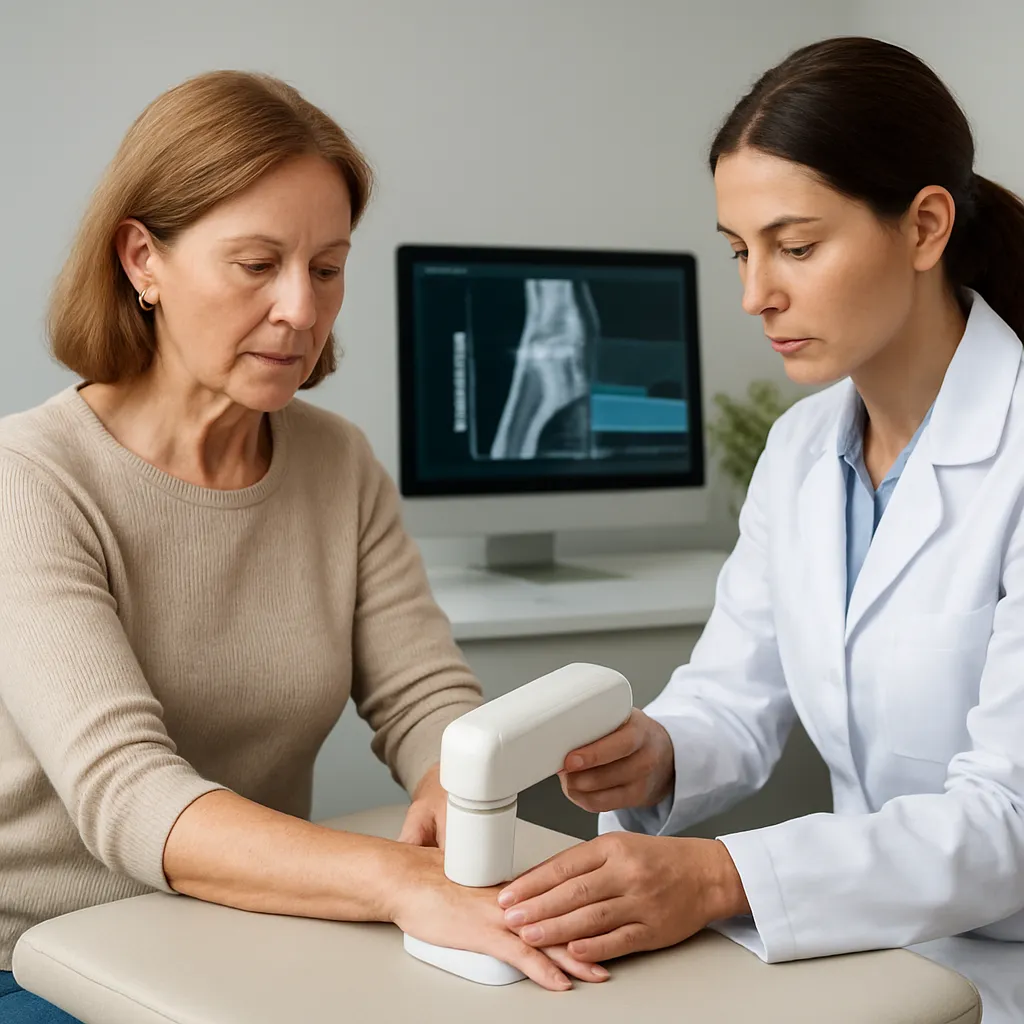
Bone density testing, most commonly performed via DXA (dual-energy X-ray absorptiometry), measures the amount of mineral in bones. The results are expressed as T-scores and Z-scores, which compare an individual’s bone density to young healthy adults and age-matched controls, respectively. A T-score ≤ –2.5 indicates osteoporosis, while a T-score between –1.0 and –2.5 is classified as osteopenia. These thresholds guide clinical decisions on prevention and therapy.
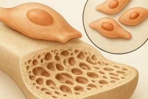
Additional modalities include quantitative computed tomography (QCT) and ultrasound-based techniques, though DXA remains the gold standard due to its high precision, low radiation exposure, and ability to assess both the spine and hip. Repeat measurements track changes over time, helping to evaluate treatment efficacy and disease progression.
- DXA: Gold standard, assesses axial skeleton.
- QCT: Volumetric analysis of trabecular bone.
- Quantitative ultrasound: Portable but less standardized.
Testing is recommended for women aged 65 and older, men aged 70 and older, and younger individuals with risk factors such as prolonged steroid use, early menopause, or a history of low-trauma fracture. Understanding the physiology of bone remodeling and the interplay of calcium, vitamin D, hormones, and mechanical stress is essential to interpreting results accurately.
Common Myths Surrounding Bone Density Screening
Despite widespread use, several misconceptions persist:
- Myth: Only elderly individuals need testing.
Fact: Adult men and women with risk factors—such as glucocorticoid therapy, rheumatoid arthritis, or family history—should be evaluated sooner.
- Myth: A single test provides lifetime assurance.
Fact: Bone density can decline rapidly, especially after surgical menopause or immobilization. Repeat scans every 1–2 years may be warranted.
- Myth: Diet alone prevents bone loss.
Fact: While adequate calcium and vitamin D are crucial, weight-bearing exercise, limiting alcohol and tobacco, and pharmacologic therapy play significant roles.
- Myth: DXA is harmful due to radiation.
Fact: Radiation exposure from a DXA scan is minimal—comparable to a day of natural background radiation—and far outweighed by its diagnostic benefit.
- Myth: High bone density guarantees no fracture risk.
Fact: Bone quality, microarchitecture, and fall risk also influence fracture susceptibility. Some patients with normal density still sustain breaks due to poor balance or underlying conditions.
By addressing these myths, patients can make informed decisions and reduce anxiety surrounding bone health assessment.
Scientific Evidence and Proven Facts
Decades of research have validated the utility of bone density testing in predicting fracture risk. A meta-analysis involving thousands of participants demonstrated that each standard deviation decrease in bone mineral density (BMD) doubles the fracture risk. Key findings include:
- Predictive Value: DXA-measured BMD at the hip and spine correlates strongly with hip, vertebral, and wrist fractures.
- Cost-Effectiveness: Early identification and treatment of osteopenia and osteoporosis reduce healthcare expenses by preventing costly hip fractures.
- Therapeutic Monitoring: Serial DXA scans gauge response to antiresorptive agents (e.g., bisphosphonates) and anabolic drugs (e.g., teriparatide).
Role of Biochemical Markers
Markers of bone turnover—such as serum C-terminal telopeptide (CTX) and procollagen type 1 N-terminal propeptide (P1NP)—complement BMD findings. High-turnover states often precede significant BMD loss, suggesting early intervention.
Integrating Clinical Risk Factors
Tools like FRAX calculate 10-year fracture probability by combining clinical factors (age, prior fracture, glucocorticoid use) with BMD data. This holistic approach informs treatment thresholds beyond density alone.
Strategies for Incorporating Testing into Practice
Effective implementation requires a systematic approach:
- Identify high-risk patients in primary care and specialty clinics, including endocrinology and geriatrics.
- Coordinate DXA scheduling, ensuring proper patient preparation—avoidance of recent contrast imaging, removal of metallic objects, and correct positioning.
- Interpret results in the context of clinical history, laboratory evaluations (e.g., serum calcium, vitamin D), and fracture risk calculators.
- Discuss findings with patients, emphasizing lifestyle modifications, fall prevention, and adherence to prescribed therapy.
Shared decision-making enhances compliance and empowers patients to participate actively in bone health management.
Emerging Trends and Future Directions
Innovations promise to refine bone health assessment:
- High-Resolution Imaging: HR-pQCT offers detailed 3D evaluation of trabecular and cortical microarchitecture, potentially improving risk stratification.
- Artificial Intelligence: AI algorithms can automate DXA interpretation, flag abnormalities, and predict fracture risk with greater accuracy.
- Biomarker Discovery: Novel serum and genetic markers may identify individuals at risk before significant BMD decline.
- Telemedicine: Remote monitoring and digital bone health platforms enable broader access, especially in underserved areas.
As research advances, integrating these tools with current screening paradigms will further personalize patient care and potentially reduce the global burden of osteoporosis-related fractures.