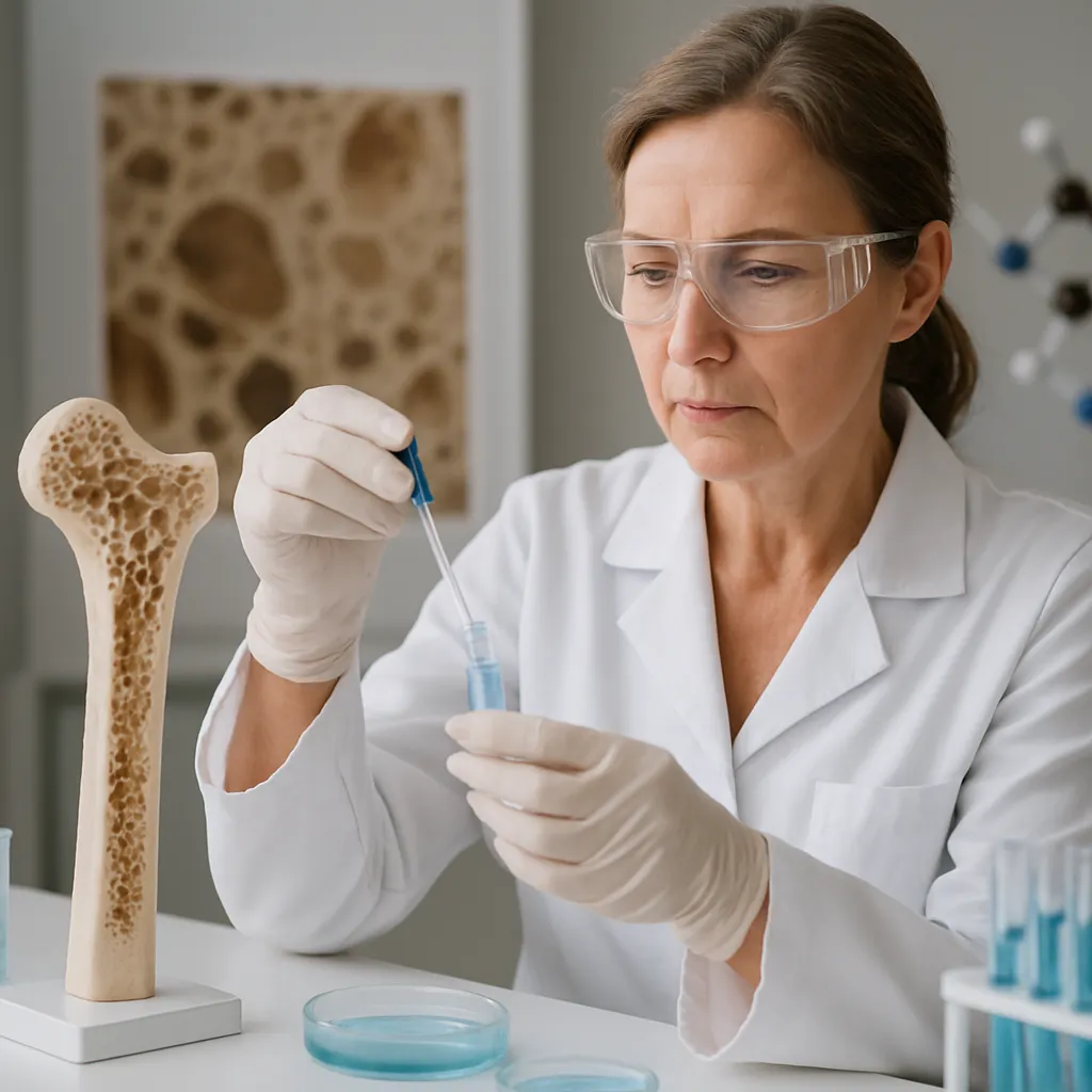
Advancements in the field of bone tissue engineering have unlocked tremendous potential for addressing complex skeletal defects. Through the integration of additive manufacturing techniques, researchers are now capable of fabricating precise three-dimensional constructs that mimic native bone architecture. This article explores the latest innovations in bioprinting strategies, delving into the essential materials, cellular components, and design parameters that propel this technology toward clinical adoption.
Overview of Bioprinting Approaches in Bone Tissue Engineering
The goal of modern bone repair is to replace or support damaged tissue by creating constructs with tailored mechanical properties, interconnected porosity, and appropriate biochemical cues. Three primary bioprinting modalities are currently under investigation:
- Inkjet-based Bioprinting: Utilizes droplet ejection to deposit cell-laden bioink in a layer-by-layer fashion. This technique offers high resolution (<50 µm) but may be limited by nozzle clogging and droplet inconsistency.
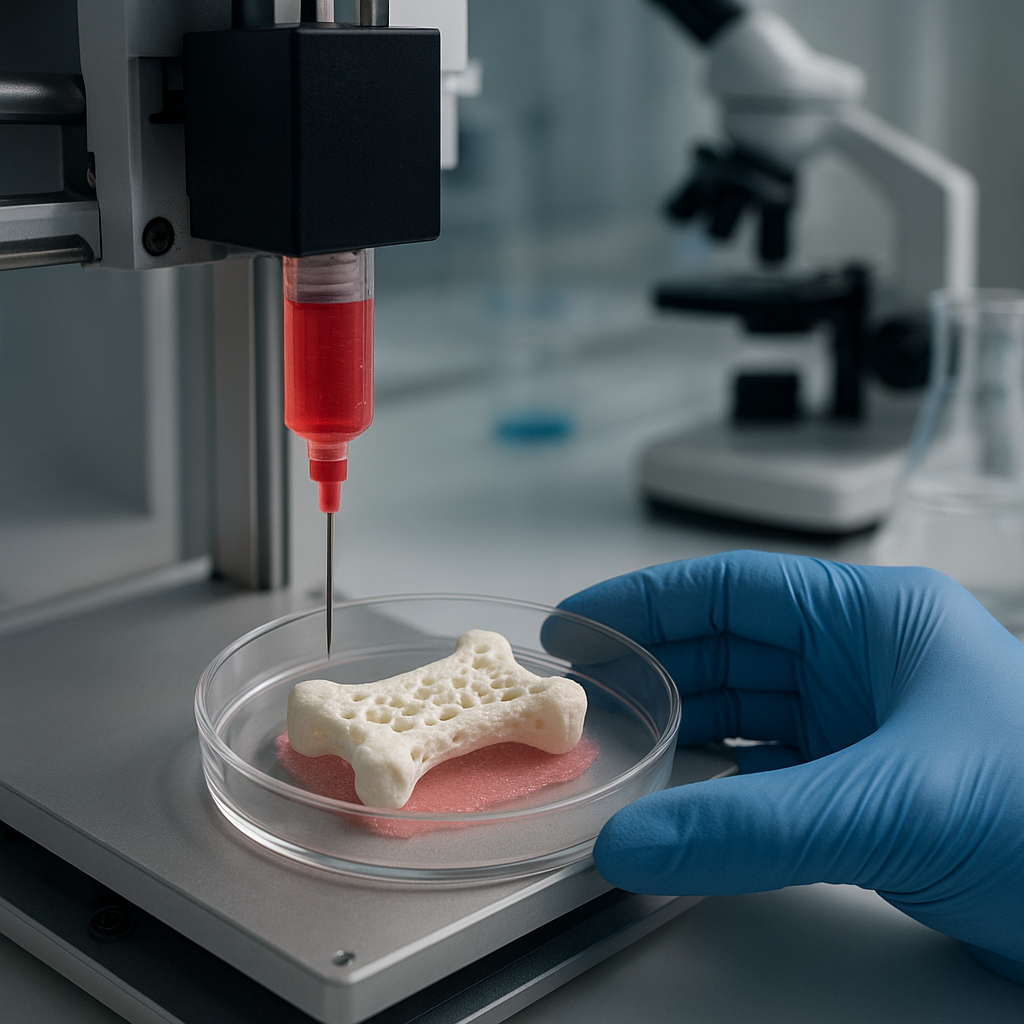
- Extrusion-based Bioprinting: Extrudes continuous filaments of hydrogels or polymer blends through a syringe-like nozzle. It accommodates a wide viscosity range, enabling integration of ceramics or nanomaterials that enhance structural integrity.
- Laser-assisted Bioprinting: Employs laser pulses to transfer microdroplets of biomaterial onto a substrate. It provides exceptional spatial accuracy and cell viability but requires complex optical setups.
Each method brings distinctive advantages in terms of resolution, printing speed, and material compatibility. Selecting the optimal technique depends on the targeted defect size, desired mechanical properties, and the biological requirements for osteogenesis.
Design Parameters and Scaffold Architecture
Scaffold geometry dictates how new bone and vasculature form within the construct. Key considerations include:
- Pore size: Ranging from 100 to 500 µm to facilitate cell infiltration and nutrient diffusion.
- Interconnectivity: Essential for promoting vascularization and waste removal.
- Mechanical strength: Matching the native bone modulus (0.1–20 GPa) to prevent stress shielding.
Advanced computer-aided design tools enable the generation of patient-specific scaffold architectures, ensuring optimal load distribution and biological integration.
Key Components: Bioinks, Scaffolds, and Cells
Successful bone bioprinting relies on the harmonious interplay of three critical elements: the choice of bioink, the supporting scaffold framework, and the seeded cell population.
Engineered Bioinks
A bioink must balance printability, biocompatibility, and mechanical stability. Typical formulations include:
- Natural polymers: Such as gelatin, collagen, and hyaluronic acid, offer cell-friendly environments but often lack robust mechanics.
- Synthetic polymers: Including polycaprolactone (PCL) and polyethylene glycol (PEG), deliver tunable degradation and strength but may require surface modification to enhance cell adhesion.
- Composite inks: Combine polymers with bioactive ceramics (e.g., hydroxyapatite) or nanomaterials like graphene oxide to boost stiffness and osteoconductivity.
Incorporation of peptides and growth factors into bioinks can further stimulate osteogenesis and guide cellular behavior.
Scaffold Materials and Fabrication
Hybrid scaffolds merge the advantages of hard and soft biomaterials. Typical strategies include:
- Core-shell structures: A rigid polymer core provides mechanical support, while a softer hydrogel shell houses cells and growth factors.
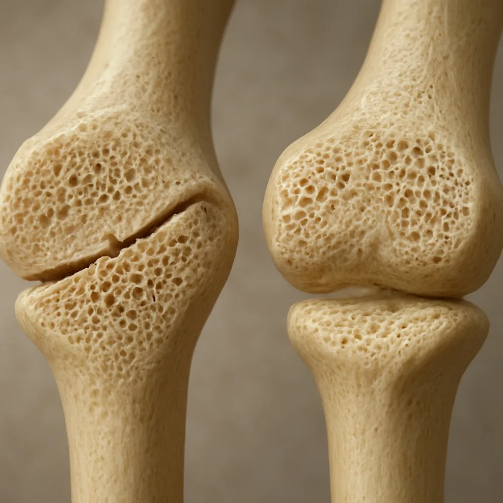
- Gradient porosity: Mimics the transition from dense cortical bone to spongy trabecular bone, facilitating efficient nutrient exchange.
- Biodegradable metals: Magnesium alloys are under exploration for temporary support, gradually resorbing as new tissue forms.
Cell Sources and Biological Factors
Choosing the right cell population is crucial for robust bone regeneration. Common cell types include:
- Mesenchymal stem cells (MSCs): Derived from bone marrow, adipose tissue, or umbilical cord–exhibit high proliferative potential and multipotency.
- Osteoblasts: Mature bone-forming cells that directly deposit mineralized matrix.
- Co-cultures with endothelial cells: Promote vascularization within printed constructs, enhancing nutrient delivery and integration.
Bioreactor systems supplying dynamic mechanical stimuli and perfusion further augment cell viability and matrix deposition.
Emerging Trends and Clinical Translation
As bioprinting moves from bench to bedside, several innovative approaches and translational challenges demand attention.
Personalized and On-Demand Constructs
Integration with medical imaging allows for the creation of patient-specific implants. High-resolution CT or MRI data guide the precise fabrication of scaffolds that conform to irregular defect geometries. This customization reduces surgery time and improves the mechanical fit, fostering faster healing.
Advanced Vascular Networks
A major hurdle remains the recreation of functional microvasculature within thick bone grafts. Promising solutions include:
- Coaxial bioprinting: Simultaneous deposition of endothelial and perivascular cells in concentric filaments to form primitive vessels.
- Sacrificial inks: Temporary carbohydrate or gelatin templates are printed and then removed, leaving perfusable channels for media flow.
- Growth factor gradients: Spatially controlled release of VEGF and BMP-2 to drive angiogenesis and bone formation in targeted regions.
Mechanical Conditioning and In Vivo Maturation
Applying cyclic compressive or tensile loads in vitro fosters extracellular matrix alignment and enhances the mechanical properties of engineered bone. Once implanted, the construct undergoes further remodeling under physiological stresses, integrating seamlessly with host tissue.
Regulatory and Manufacturing Considerations
Scaling up production for clinical use introduces regulatory complexities. Key factors include:
- Material traceability and sterility assurance.
- Standardized protocols for cell sourcing and handling.
- Quality control measures to verify mechanical performance and cell viability batch-to-batch.
Collaborations between engineering teams, biologists, and regulatory experts are essential to streamline the path from laboratory innovation to routine medical practice.
Future Directions in Bone Bioprinting
Looking ahead, the next generation of bone constructs will likely feature integrated sensors for real-time health monitoring, smart materials that respond to biochemical signals, and AI-driven design algorithms that optimize scaffold architecture. Advances in genetic engineering may yield stem cells programmed to secrete enhanced osteoinductive factors, while modular printing platforms could allow simultaneous deposition of hard and soft tissues, recreating the bone–cartilage interface for joint repair.
As these technologies converge, the vision of fully functional, lab-grown bone implants moves ever closer to reality. The continued refinement of regeneration strategies, combined with a deeper understanding of bone biomechanics and cellular interactions, promises to transform the landscape of orthopedic medicine and offer new hope for patients with challenging skeletal defects.