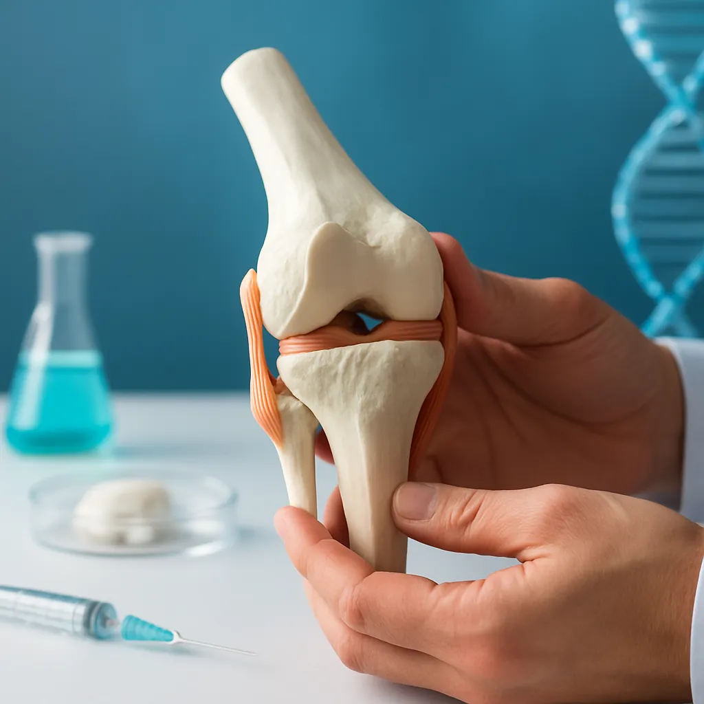





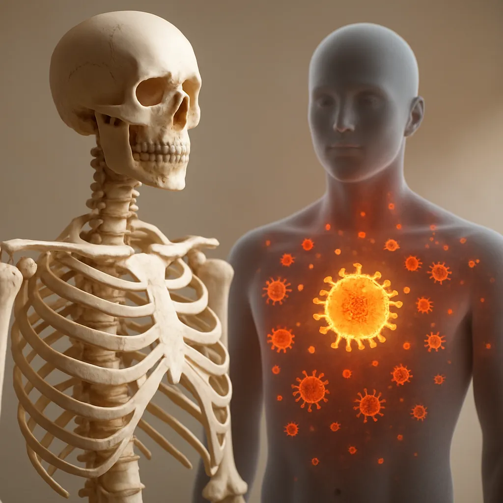
The intricate relationship between the skeletal system and the immune network has garnered significant attention as scientists uncover how bones actively regulate immune cell development and function. Far from serving solely as a mechanical framework, bone tissue provides a rich microenvironment in which hematopoietic cells differentiate, mature, and respond to challenges. Understanding this bidirectional communication sheds light on mechanisms underlying diseases such as osteoporosis, rheumatoid arthritis, and bone marrow failure syndromes, as well as guides the design of novel therapies.
Bone as a Dynamic Organ System
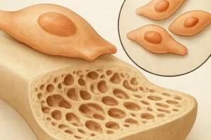
Contrary to the outdated view of bone as a static structure, it is a highly metabolic organ undergoing continual bone remodeling. This process involves the balanced activity of two specialized cell types: osteoclasts, which resorb old bone matrix, and osteoblasts, which synthesize new matrix. Together, they orchestrate maintenance of bone density and integrity. Key factors regulating their actions include hormonal signals (e.g., parathyroid hormone, estrogen), mechanical loading, and molecular cues from the immune system.
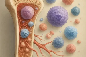
Within the rigid matrix, the bone marrow cavity houses a complex milieu where mesenchymal stromal cells, endothelial cells, and hematopoietic progenitors interact. Here, the compartmentalization into endosteal, perivascular, and central niches fosters distinct stages of blood cell maturation. Importantly, the secretome of stromal cells—encompassing growth factors and cytokines—influences not only hematopoiesis but also the activity of bone-resorbing and bone-forming cells.
Mechanical stresses, transmitted through weight-bearing and microfractures, trigger mechanotransduction pathways that modulate gene expression in bone cells. This, in turn, can indirectly alter immune cell egress and retention within the marrow. Thus, skeletal loading has systemic immunological consequences, highlighting a feedback loop between physical activity and immune competence.
Immune Cells within the Bone Marrow Microenvironment
The bone marrow is the principal site of hematopoiesis, generating all blood cells, including lymphocytes, monocytes, and granulocytes. As immune cells mature, they rely on interactions with stromal elements and extracellular matrix components. For example, the chemokine CXCL12, produced by stromal cells, retains hematopoietic stem cells (HSCs) in niches until differentiation cues are received.
Key immune populations within the marrow include:
- Macrophages: These phagocytes clear apoptotic cells and secrete factors that influence both HSC quiescence and activation of osteoblasts.
- Dendritic cells: Present antigens to T cells but also produce RANKL and M-CSF, which can stimulate osteoclast differentiation.
- Regulatory T cells (Tregs): They maintain local immune tolerance and secrete anti-inflammatory cytokines (IL-10, TGF-β) that suppress excessive bone resorption.
- Neutrophils: Although short-lived, they release proteases and reactive species that can modulate niche integrity during infection or injury.
During systemic infection or injury, emergency hematopoiesis in the marrow accelerates, diverting resources toward rapid innate cell production. This shift often results in temporary alterations in bone turnover, as inflammatory mediators induce osteoclastogenesis to release minerals and growth factors stored in bone, facilitating immune cell expansion.
Cytokines and Signaling Pathways in Osteoimmunology
Communication between bone and immune systems is largely mediated by cytokines and growth factors. Critical molecular players include:
- RANK/RANKL/OPG axis: RANKL (Receptor Activator of Nuclear factor κB Ligand), expressed by osteoblasts and activated T cells, binds RANK on osteoclast precursors to promote their maturation. Osteoprotegerin (OPG) acts as a decoy receptor, limiting RANKL’s effects and acting as a brake on resorption.
- Interleukins: IL-1, IL-6, and TNF-α produced by macrophages and T cells enhance osteoclast differentiation and activity, linking inflammation with bone loss.
- Wnt signaling: This pathway governs osteoblast proliferation and survival. Wnt antagonists such as sclerostin, secreted by osteocytes, inhibit bone formation and can be modulated by inflammatory stimuli.
- Interferon-γ (IFN-γ): While it promotes macrophage activation and pathogen clearance, IFN-γ also inhibits osteoclastogenesis, exemplifying the nuanced crosstalk between immunity and skeletal homeostasis.
In autoimmune conditions like rheumatoid arthritis, dysregulated cytokine production sustains a vicious cycle of synovial inflammation and bone erosion. Elevated levels of TNF-α and IL-17 drive osteoclast activation, while insufficient counter-regulatory signals fail to curb destruction. Such insights have transformed clinical practice, with biologic agents targeting specific cytokines to protect joints and preserve skeletal integrity.
Clinical Implications and Therapeutic Perspectives
The burgeoning field of osteoimmunology informs a variety of medical interventions. Strategies include:
- Biologics against TNF-α, IL-6R, and IL-17: These agents mitigate excessive bone resorption in autoimmune disorders and alleviate systemic bone loss.
- Denosumab: A monoclonal antibody mimicking OPG, it inhibits RANKL, reducing osteoclast activity and improving bone density in osteoporosis patients.
- Emerging modulators of sclerostin: By stimulating Wnt signaling, these therapies enhance osteoblast function and are under investigation for fracture prevention.
- Cell therapies: Infusion of mesenchymal stem cells or regulatory T cells shows promise in restoring tolerance and supporting bone regeneration in post-transplant and autoimmune settings.
In oncology, understanding how tumor cells hijack the bone marrow niche to evade immune surveillance has opened avenues for combining immunotherapies with bone-targeted agents. Blocking myeloid-derived suppressor cell expansion or reprogramming macrophages within bone metastatic sites holds potential to improve outcomes in multiple myeloma and breast cancer metastases.
Future research aims to identify novel therapeutic targets by mapping the molecular dialogue between bone matrix components and immune receptors. Single-cell transcriptomics and advanced imaging techniques will elucidate niche heterogeneity and reveal intervention points to enhance marrow resilience. As we unravel the complexities of bone–immune interactions, personalized approaches to maintain skeletal health and optimize immune function will become increasingly feasible.