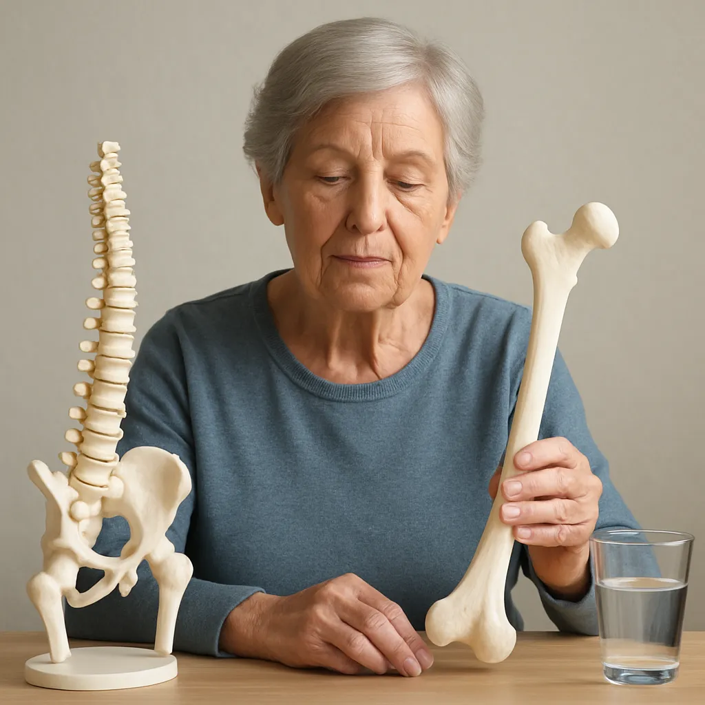
The pelvic bone, a complex structure located at the base of the spine, plays a crucial role in the human body. It serves as a foundation for the spine, supports the weight of the upper body, and provides attachment points for various muscles and ligaments. Understanding the anatomy and function of the pelvic bone is essential for comprehending its significance in both movement and overall health. This article will delve into the intricate details of the pelvic bone’s structure and its vital functions in the human body.
Anatomy of the Pelvic Bone
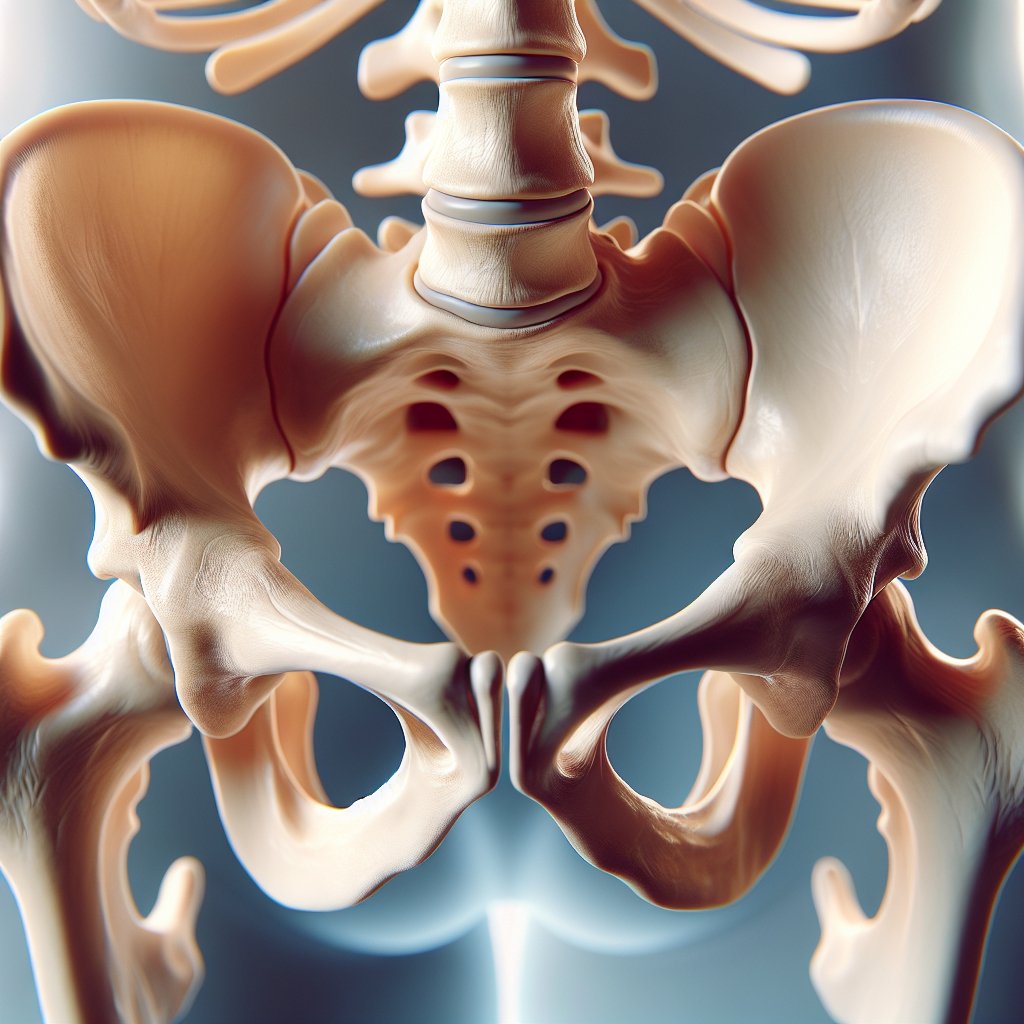
The pelvic bone, also known as the pelvis, is composed of several key components that work together to form a sturdy yet flexible structure. It is primarily made up of three bones: the ilium, ischium, and pubis, which fuse together during adolescence to create a single bone known as the innominate bone. The pelvis can be divided into two main parts: the greater (or false) pelvis and the lesser (or true) pelvis.
Components of the Pelvic Bone
- Ilium: The largest part of the pelvis, the ilium is the broad, flaring portion that forms the upper part of the pelvic bone. It provides attachment points for various muscles, including those involved in hip movement.
- Ischium: Located at the lower and back part of the pelvis, the ischium is the bone that you sit on. It provides structural support and serves as an attachment point for muscles of the thigh and hip.
- Pubis: The pubis is the front portion of the pelvis and is connected to the other side of the pubis by the pubic symphysis, a cartilaginous joint. It plays a role in stabilizing the pelvis during movement.
In addition to these three main bones, the pelvic bone also includes several important features:
- Acetabulum: This is the socket of the hip joint, where the head of the femur (thigh bone) fits, allowing for a wide range of motion in the hip.
- Pelvic inlet and outlet: The pelvic inlet is the upper opening of the pelvis, while the pelvic outlet is the lower opening. These openings are crucial for childbirth, as they determine the size and shape of the birth canal.
- Obturator foramen: This is a large opening in the pelvis that allows for the passage of nerves and blood vessels to the lower limbs.
Pelvic Girdle and Its Joints
The pelvic bone is part of the pelvic girdle, which connects the spine to the lower limbs. The pelvic girdle consists of the two innominate bones (one on each side) and the sacrum, which is the triangular bone at the base of the spine. The sacroiliac joints connect the sacrum to the ilium, providing stability and support during movement.
Another important joint in the pelvis is the pubic symphysis, which connects the two pubic bones at the front of the pelvis. This joint allows for slight movement, which is particularly important during childbirth, as it can help accommodate the passage of the baby through the birth canal.
Function of the Pelvic Bone
The pelvic bone serves several essential functions that are vital for human movement, stability, and overall health. Its design allows for a combination of strength and flexibility, making it an integral part of the body’s musculoskeletal system.
Support and Weight Bearing
One of the primary functions of the pelvic bone is to support the weight of the upper body when standing, walking, or sitting. The pelvis acts as a stable base for the spine, distributing weight evenly across the lower limbs. This weight-bearing function is crucial for maintaining balance and posture, especially during activities that involve movement or physical exertion.
Facilitating Movement
The pelvic bone plays a significant role in facilitating movement, particularly in the hips and legs. The acetabulum allows for a wide range of motion in the hip joint, enabling activities such as walking, running, and jumping. The muscles that attach to the pelvic bone, including the gluteal muscles and hip flexors, are essential for generating movement and providing stability during physical activities.
Childbirth
In females, the pelvic bone has a unique function related to childbirth. The shape and size of the pelvis determine the dimensions of the birth canal, which is critical for the safe delivery of a baby. During pregnancy, hormonal changes cause the ligaments in the pelvis to relax, allowing for greater flexibility and expansion during labor. This adaptation is vital for accommodating the baby’s passage through the birth canal.
Protection of Internal Organs
The pelvic bone also serves a protective function, safeguarding the internal organs located within the pelvic cavity. These organs include the bladder, reproductive organs, and parts of the digestive system. The bony structure of the pelvis provides a shield against external trauma, helping to prevent injury to these vital organs.
Posture and Stability
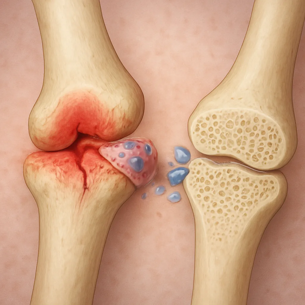
Proper alignment and stability of the pelvis are essential for maintaining good posture. The pelvic bone acts as a keystone in the body’s structure, influencing the alignment of the spine and lower limbs. Imbalances or misalignments in the pelvis can lead to various musculoskeletal issues, including lower back pain, hip pain, and knee problems. Therefore, maintaining pelvic health is crucial for overall well-being.
Conclusion
The pelvic bone is a remarkable structure that plays a multifaceted role in the human body. Its intricate anatomy, comprising the ilium, ischium, and pubis, allows for a combination of strength and flexibility essential for movement, stability, and protection of internal organs. Understanding the anatomy and function of the pelvic bone is vital for appreciating its significance in daily activities and overall health. Whether it is supporting the weight of the body, facilitating movement, or playing a crucial role in childbirth, the pelvic bone is an indispensable component of the human musculoskeletal system.