The intricate interplay between the human skeleton and the musculature system underpins our ability to move, bear loads, and maintain overall health. As scientific inquiry delves deeper into the mechanisms that link bone density with muscle strength, clinicians and researchers uncover strategies to optimize skeletal integrity, prevent degeneration, and enhance quality of life. This article explores fundamental concepts of bone-muscle interactions, examines key determinants of structural and functional adaptation, and offers practical insights for medical and athletic contexts.
The Physiological Basis of Bone-Muscle Interactions
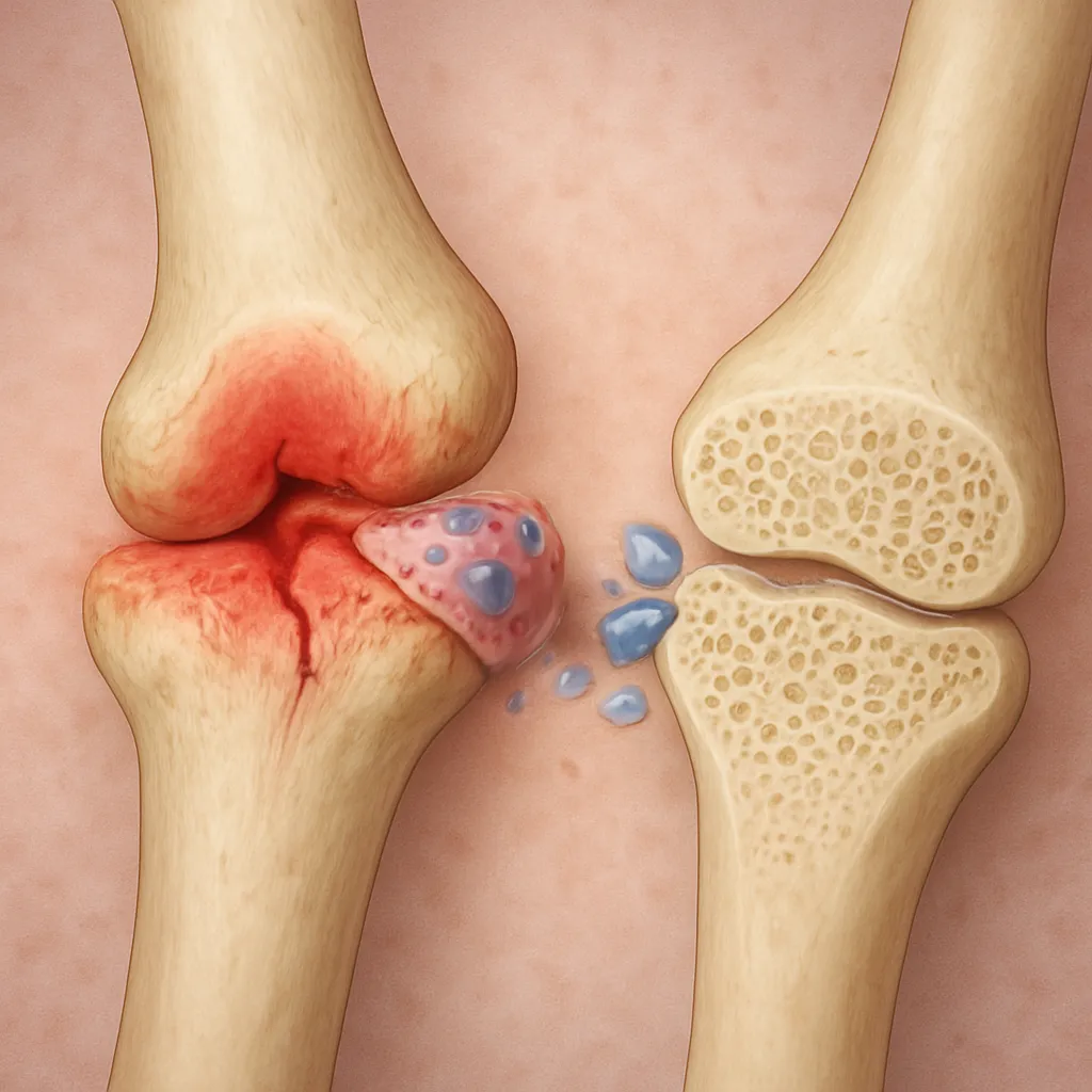
At the core of the bone-muscle relationship lies a dynamic process of remodeling, in which bone tissue continuously adapts to mechanical demands imposed by muscle contraction. According to Wolff’s law and the mechanostat theory, bones sense strain and adjust their architecture to maintain optimal strength. When muscles contract, they generate forces that are transmitted to bones, stimulating osteoblast activity and promoting new bone formation. Conversely, insufficient loading triggers osteoclast-mediated resorption, leading to structural weakening.
Bone Remodeling and Cellular Players
- Osteoblasts: Cells responsible for synthesizing the organic matrix—primarily collagen—and facilitating mineral deposition.
- Osteoclasts: Multinucleated cells that resorb mineralized bone, releasing calcium into the bloodstream.
- Osteocytes: Mature bone cells embedded within the matrix, acting as mechanosensors that regulate remodeling rates.
These cellular activities are tightly regulated by signaling molecules, such as RANKL, OPG, and various growth factors, which respond to mechanical cues and systemic influences. The balance between resorption and formation determines the overall peak bone mass attained during early adulthood and the subsequent trajectory of bone health throughout life.
Muscular Forces and Skeletal Adaptation
Muscle fibers contract through excitation-contraction coupling, generating tensile forces transmitted via tendons to the skeletal system. The magnitude, frequency, and direction of these forces shape bone geometry and density. High-impact activities—such as jumping or sprinting—induce rapid loading rates that challenge bone’s structural capacity, stimulating adaptive responses. Meanwhile, low-intensity, prolonged activity yields less potent osteogenic stimuli, underscoring the importance of targeted interventions to preserve skeletal health.
Factors Influencing Bone Density and Muscle Strength
Multiple determinants converge to influence both bone mineralization and muscular performance. Genetic predispositions set the baseline for potential strength and density, while environmental factors and lifestyle choices modulate outcomes appreciably. Understanding these variables is crucial for designing preventive and therapeutic approaches.
Nutrition and Metabolic Support
- Calcium metabolism: Adequate calcium intake is essential for mineral deposition; vitamin D facilitates intestinal absorption and supports endocrine pathways that regulate bone remodeling.
- Protein consumption: Amino acids supply the building blocks for muscle contractile proteins and bone matrix components.
- Trace minerals: Magnesium, phosphorus, and zinc play critical roles in enzymatic processes related to both bone mineral density and muscle function.
Hormonal Regulation
Hormones orchestrate the balance between anabolism and catabolism in bone and muscle tissues. For example, sex steroids—estrogen and testosterone—promote osteoblast activity and inhibit bone resorption. Hormonal regulation also extends to growth hormone, insulin-like growth factor-1 (IGF-1), and cortisol, each exerting significant effects on tissue turnover. Dysregulation of these hormones can precipitate conditions such as osteoporosis or sarcopenia, highlighting the need for endocrine assessment in at-risk populations.
Physical Activity and Mechanical Loading
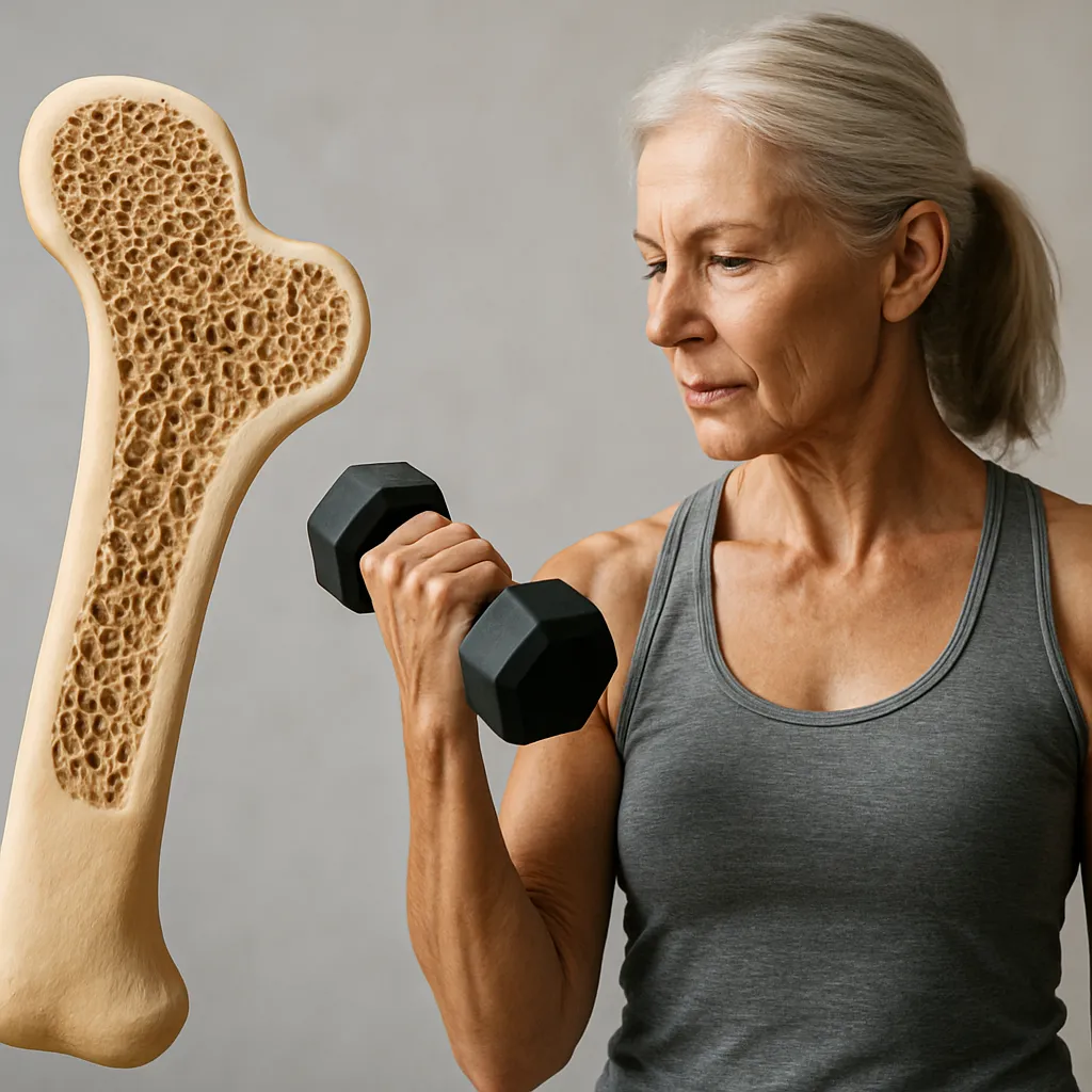
- Resistance training: Progressive overload through weightlifting or bodyweight exercises enhances muscle cross-sectional area and exerts targeted stress on bones, promoting site-specific mineral accrual.
- Aerobic exercise: High-impact running and plyometric drills deliver mechanical loading patterns that stimulate periosteal bone formation.
- Immobilization: Extended bed rest or microgravity exposure leads to rapid declines in both muscle strength and bone density, underscoring the principle of “use it or lose it.”
Age, Sex, and Genetic Factors
Peak bone mass is typically attained by the third decade of life, after which gradual remodeling imbalances may favor resorption. Aging also coincides with reductions in muscle fiber number and contractile function. Genetic polymorphisms influence collagen structure, vitamin D receptor function, and myostatin activity, thereby shaping individual variability in adaptation. Sex-specific differences—such as the accelerated bone loss observed in postmenopausal women—necessitate tailored monitoring and intervention strategies.
Practical Implications for Clinical Practice and Training
Translating the insights from bone-muscle research into actionable protocols can improve outcomes in diverse settings, from rehabilitation clinics to athletic facilities. By integrating assessment tools, nutritional guidance, and exercise prescription, practitioners can foster robust musculoskeletal health across the lifespan.
Clinical Assessment and Monitoring
- Dual-energy X-ray absorptiometry (DXA): Gold-standard technique for measuring bone density at clinically relevant sites such as the hip and spine.
- Quantitative ultrasound: Portable option for evaluating peripheral bone properties.
- Isokinetic dynamometry: Provides objective measures of muscle strength and power output.
- Biochemical markers: Serum levels of alkaline phosphatase, collagen cross-links, and vitamin D metabolites inform remodeling rates.
Exercise Prescription Guidelines
Evidence supports the use of targeted exercise regimens to maximize concurrent gains in bone and muscle. Key principles include:
- Progressive overload: Incrementally increasing resistance to challenge both muscle fibers and bone tissue.
- Specificity: Selecting movements that reproduce functional loading patterns, such as squats, deadlifts, and lunges, to benefit the lumbar spine and lower limbs.
- Frequency and intensity: Performing weight-bearing exercise sessions at least three times per week, with moderate to high intensity, to optimize osteogenic and myogenic responses.
- Variety: Incorporating dynamic, multidirectional activities to stimulate adaptation across different anatomical sites.
Nutritional and Lifestyle Strategies
Dietary optimization complements mechanical interventions by supplying essential substrates and cofactors. Recommended practices include:
- A balanced diet with 1.2 to 1.5 grams of protein per kilogram of body weight to support muscle hypertrophy and bone matrix synthesis.
- Calcium intake of at least 1,000 to 1,200 mg per day, paired with sufficient vitamin D exposure through sunlight and/or supplementation.
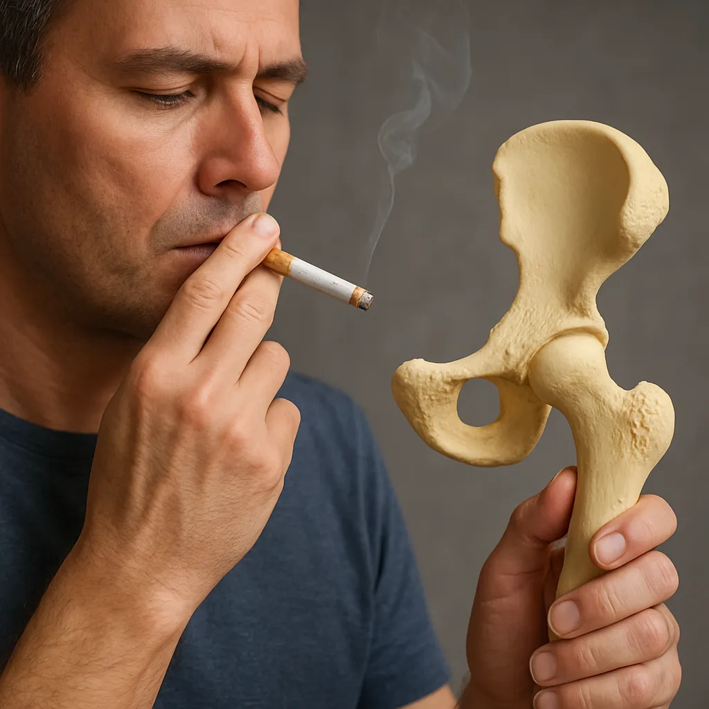
- Limiting alcohol consumption and avoiding tobacco use, both of which impair bone formation and muscle repair.
- Regular screening for hormonal imbalances and early treatment of thyroid or endocrine disorders that can disrupt calcium metabolism and tissue turnover.
By embracing a holistic approach that integrates biomechanics, nutrition, and personalized exercise, healthcare providers can help individuals achieve and maintain optimal musculoskeletal function. The synergy between muscle contractions and bone adaptation underscores a fundamental principle: strong muscles and dense bones are inextricably linked, and interventions that target one often benefit the other. Continued research into cellular signaling pathways, novel training modalities, and genetic markers will refine strategies to prevent degenerative conditions and enhance performance across all age groups.