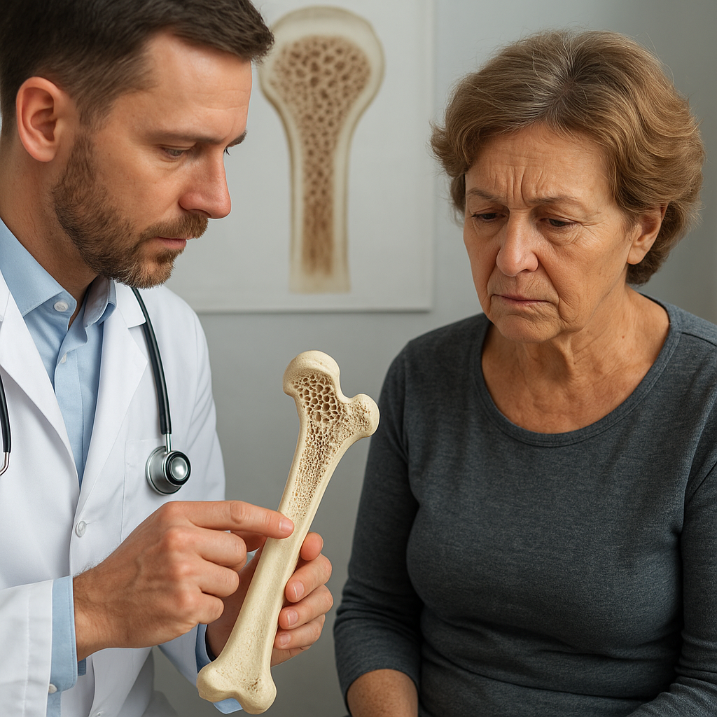
Bone allograft preservation has undergone significant evolution, driven by the need to maintain the structural integrity and biological activity of donor tissue. Researchers and clinicians have collaborated to develop advanced protocols that ensure optimal outcomes in orthopaedic and reconstructive surgery. This article explores various facets of bone allograft preservation, highlighting cutting-edge techniques and materials.
Preservation Techniques in Bone Allografting
Effective preservation methods are fundamental to maintaining the mechanical strength and biocompatibility of bone grafts. Traditional protocols have expanded to embrace innovative approaches designed to reduce immunogenicity while enhancing osteoinductive properties.
Fresh-Frozen Preservation
Fresh-frozen bone allografts remain a mainstay in clinical practice due to their ability to retain a high degree of native matrix proteins and growth factors. The tissue is harvested aseptically, tested for pathogens, and rapidly frozen at temperatures below –80°C. Advantages include:
- Retention of extracellular matrix architecture
- Preservation of collagen and non-collagenous proteins
- Improved osteoinduction compared to freeze-dried grafts
However, freeze-thaw cycles can lead to formation of ice crystals that disrupt cellular structures. Use of cryoprotectants such as glycerol or dimethyl sulfoxide (DMSO) helps mitigate ice damage by lowering the freezing point and stabilizing membranes.
Freeze-Drying (Lyophilization)
Lyophilization removes water content through sublimation under vacuum, producing a stable, dry graft that can be stored at ambient temperature. Key benefits include:
- Extended shelf life without refrigeration
- Reduced risk of microbial growth
- Maintained structural geometry
Despite these advantages, the process can denature delicate proteins responsible for osteogenesis. To counter this, optimized freezing rates and secondary drying steps have been introduced, preserving higher levels of bone morphogenetic proteins (BMPs).
Cryopreservation with Controlled-Rate Freezing
Controlled-rate freezing offers a precise method to freeze bone tissue at a regulated speed, preventing rapid ice crystal formation. This approach involves:
- Programmable freezing devices
- Stepwise temperature reduction (e.g., –1°C/min)
- Integration of cryoprotective agents
Research indicates that combining cryopreservation with perfusion of protective solutions enhances viability of osteocytes and preserves the anatomical microarchitecture critical for load-bearing applications.
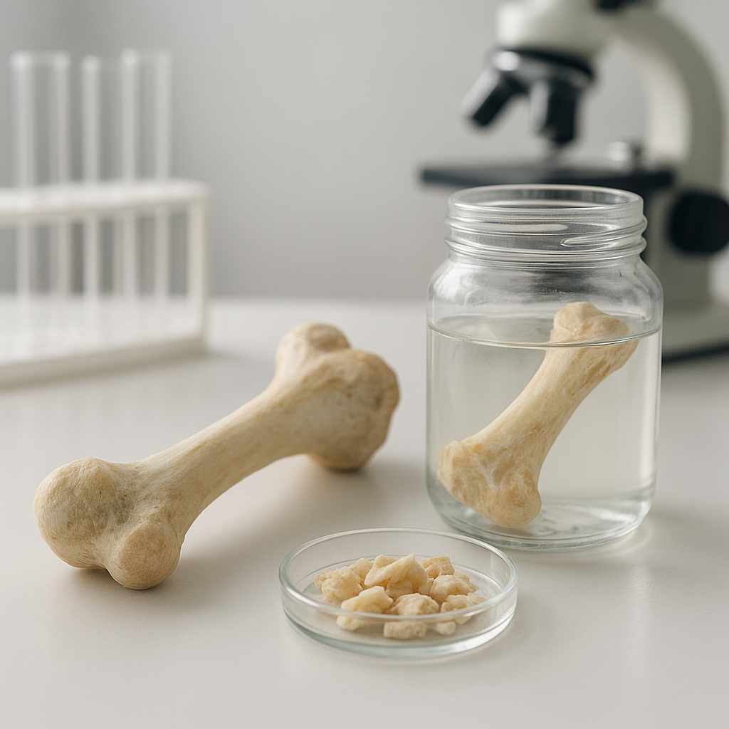
Innovations in Decellularization and Sterilization
Removing cellular components from allograft bone reduces the risk of immune rejection. Decellularization must balance thorough cell removal with preservation of the collagenous scaffold and native growth factors.
Chemical Decellularization Protocols
Chemical methods employ detergents, acids, or enzymes to solubilize cell membranes and intracellular debris. Common agents include:
- Sodium dodecyl sulfate (SDS)
- Triton X-100
- Trypsin-EDTA
Recent protocols utilize sequential treatments—first with hypotonic buffers to induce cell lysis, followed by enzymatic digestion and thorough washing steps. Application of ultrasonication has proven effective in further dislodging residual cellular fragments without compromising scaffold integrity.
Supercritical CO2 Sterilization
Supercritical carbon dioxide represents a novel, low-temperature alternative to gamma irradiation and ethylene oxide. Characteristics include:
- Antimicrobial efficacy through lipid extraction
- Minimal alteration of mechanical properties
- No toxic residues
This technique is particularly attractive for preserving sensitive proteins and ensuring optimal scaffold performance. Studies have shown that supercritical CO2-treated allografts retain >90% of their compressive strength and exhibit negligible changes in collagen crosslinking.
Optimized Gamma Irradiation
Gamma irradiation has long been a gold standard for terminal sterilization. However, high doses (>25 kGy) can lead to brittle grafts. To minimize damage, practitioners now:
- Apply fractionated irradiation doses
- Use radical scavengers such as mannitol or trehalose
- Combine irradiation with low-temperature processing
These improvements reduce free radical formation, preserving the biomechanical performance and structural proteins within the bone matrix.
Emerging Biomaterials and Composite Scaffolds
Synthetic and hybrid scaffolds are being developed to augment allografts, promoting faster integration and increased mechanical resilience. Composite materials can deliver controlled release of growth factors and antibiotics, supporting both regeneration and infection control.
Calcium Phosphate Ceramics
Hydroxyapatite (HA) and beta-tricalcium phosphate (β-TCP) have chemical similarity to bone mineral, offering excellent osteoconductivity. Composite formulations involve:
- Adding HA nanoparticles to freeze-dried grafts for enhanced porosity
- Incorporating β-TCP granules in cryopreserved blocks
- Embedding antibiotic-loaded ceramics to prevent post-operative infections
These hybrids improve cell attachment and new bone formation while maintaining mechanical integrity suitable for load-bearing sites.
Polymeric Hydrogels and Injectable Scaffolds
Injectable systems based on alginate, chitosan, or polyethylene glycol (PEG) allow minimally invasive delivery of graft composites. Innovation in this area includes:
- Photo-crosslinkable PEG hydrogels that solidify in situ
- Thermo-responsive polymers that gel at body temperature
- Peptide-functionalized matrices promoting specific cell interactions
When combined with allograft particles, these matrices fill irregular defects and provide a sustained release of bioactive cues, enhancing cellular infiltration and vascularization.
Nanocomposite Scaffolds
Nanoscale reinforcement with silica, graphene oxide, or titanium dioxide can elevate the mechanical performance of grafts. Key advances include:
- Electrospun nanofibers that mimic collagen fibrils
- Surface functionalization with RGD peptides for improved cell adhesion
- Hybridization with magnetic nanoparticles for targeted therapy
Such materials leverage the high surface area-to-volume ratio, promoting protein adsorption and accelerating the natural bone remodeling cycle.
Regulatory and Clinical Considerations
Successful translation of preservation innovations requires rigorous compliance with regulatory standards. Tissue banks must adhere to Good Tissue Practices (GTP) and implement validated quality control assays for:
- Microbial bioburden assessment
- Residual chemical quantification
- Mechanical testing under simulated physiological loads
Clinical adoption hinges on demonstrable safety and efficacy through randomized trials, long-term follow-up studies, and post-market surveillance. Surgeons play a crucial role in selecting the most appropriate allograft type based on defect size, anatomical location, and patient-specific factors such as bone quality and comorbidities.
The convergence of advanced preservation methods, novel decellularization strategies, and engineered composite scaffolds is poised to transform the landscape of bone allografting. Ongoing research continues to refine protocols, ensuring that future grafts deliver superior performance and predictable outcomes in bone repair and regeneration.