A bone scan is a diagnostic imaging technique that plays a crucial role in the evaluation of various bone conditions. This non-invasive procedure utilizes a small amount of radioactive material to detect abnormalities in the bones, making it an essential tool for healthcare providers. Understanding what a bone scan entails and the circumstances under which it is needed can help patients make informed decisions about their health and treatment options.
Understanding Bone Scans
A bone scan, also known as a skeletal scintigraphy, is a nuclear imaging technique that provides valuable information about the metabolic activity of bones. Unlike traditional X-rays, which primarily show the structure of bones, a bone scan reveals how well the bones are functioning. This is particularly useful for identifying conditions that may not be visible on standard imaging tests.
The procedure involves the injection of a radioactive tracer, typically technetium-99m, into a vein. This tracer is attracted to areas of high bone activity, such as inflammation, infection, or tumors. After allowing time for the tracer to circulate and accumulate in the bones, a special camera is used to capture images of the skeletal system. The resulting images can highlight areas of abnormal bone metabolism, providing insights into various medical conditions.
How a Bone Scan Works
The process of a bone scan can be broken down into several key steps:
- Preparation: Patients may be advised to drink plenty of fluids before the scan to help flush out the tracer from the body after the procedure.
- Injection: A healthcare professional administers the radioactive tracer through an intravenous (IV) line. The amount of radiation used is minimal and considered safe.
- Waiting Period: After the injection, patients typically wait for two to three hours to allow the tracer to accumulate in the bones.
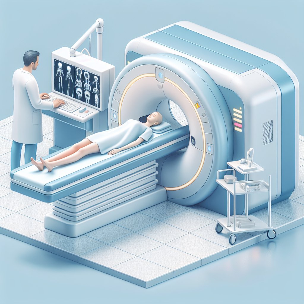
- Imaging: Patients lie down on a scanning table while a gamma camera captures images of the bones. The entire process usually takes about 30 to 60 minutes.
Once the images are obtained, a radiologist interprets the results and provides a report to the referring physician. This report can help guide further diagnostic testing or treatment options based on the findings.
Indications for a Bone Scan
Bone scans are utilized in a variety of clinical scenarios. Understanding when a bone scan is needed can help patients and healthcare providers make timely and informed decisions regarding diagnosis and treatment.
1. Detecting Bone Metastases
One of the primary uses of a bone scan is to detect metastatic cancer that has spread to the bones. Many types of cancer, including breast, prostate, and lung cancer, can metastasize to the skeletal system. A bone scan can identify these areas of metastasis, allowing for early intervention and treatment planning.
2. Evaluating Bone Pain
Patients experiencing unexplained bone pain may benefit from a bone scan. The imaging can help identify the underlying cause of the pain, such as fractures, infections, or inflammatory conditions. By pinpointing the source of discomfort, healthcare providers can tailor treatment strategies to address the specific issue.
3. Diagnosing Infections
Bone infections, or osteomyelitis, can be challenging to diagnose using conventional imaging methods. A bone scan can reveal areas of increased metabolic activity associated with infection, aiding in the diagnosis and subsequent treatment of the condition.
4. Assessing Arthritis and Other Inflammatory Conditions
Bone scans can also be useful in evaluating inflammatory conditions such as rheumatoid arthritis or psoriatic arthritis. By highlighting areas of inflammation in the bones and joints, healthcare providers can better understand the extent of the disease and monitor treatment effectiveness.
5. Monitoring Treatment Response
For patients undergoing treatment for bone-related conditions, such as cancer or arthritis, bone scans can be employed to monitor the effectiveness of therapy. Changes in the metabolic activity of the bones can indicate whether the treatment is working or if adjustments are necessary.
Benefits and Risks of Bone Scans
Like any medical procedure, bone scans come with their own set of benefits and risks. Understanding these factors can help patients weigh the pros and cons of undergoing the procedure.
Benefits
- Non-invasive: Bone scans are generally non-invasive and do not require any surgical procedures.
- Comprehensive Information: The scans provide valuable insights into bone metabolism, which can aid in diagnosing various conditions.
- Early Detection: Bone scans can detect abnormalities before they become apparent on X-rays, allowing for earlier intervention.
- Guiding Treatment: The results can help guide treatment decisions and monitor the effectiveness of ongoing therapies.
Risks
- Radiation Exposure: Although the amount of radiation used in a bone scan is minimal, there is still a small risk associated with exposure to radioactive materials.
- Allergic Reactions: In rare cases, patients may experience allergic reactions to the radioactive tracer.
- False Positives: Bone scans can sometimes yield false-positive results, leading to unnecessary anxiety or further testing.
Conclusion
A bone scan is a valuable diagnostic tool that provides critical information about bone health and metabolism. By understanding what a bone scan is and when it is needed, patients can engage in informed discussions with their healthcare providers about their symptoms and treatment options. Whether it is for detecting cancer metastases, evaluating unexplained bone pain, or monitoring treatment response, bone scans play a significant role in modern medicine. As with any medical procedure, it is essential to consider the benefits and risks, ensuring that patients receive the most appropriate care for their individual needs.