Bone is a dynamic tissue that continuously adapts to the demands placed upon it by mechanical forces. This adaptability is crucial not only for maintaining structural integrity but also for ensuring metabolic functions and overall skeletal health. Understanding the complex interplay between mechanical stress and bone physiology is vital for devising new therapies and optimizing physical activity regimens to prevent and treat various bone disorders.
Mechanisms of Bone Adaptation
The skeleton responds to physical stress through a finely tuned cellular network that senses and translates mechanical cues into biochemical signals. This process, known as mechanotransduction, involves several distinct phases: stress detection, signal propagation, and effector response. Initially, forces deform the extracellular matrix, generating interstitial fluid flow within the lacuno-canalicular system. Mechanosensitive cells then detect these changes and convert them into intracellular second messengers.
Cellular Players in Adaptation
- Osteocytes: As the most abundant bone cells, they serve as primary mechanosensors. Situated in lacunae and connected via canaliculi, osteocytes sense fluid shear stress and orchestrate recruitment of bone-forming or bone-resorbing cells.
- Osteoblasts: These matrix-producing cells originate from mesenchymal stem cells and are responsible for new bone synthesis. They express collagen type I and alkaline phosphatase and regulate mineral deposition in response to mechanical stimuli.
- Osteoclasts: Derived from hematopoietic precursors, these multinucleated cells dissolve mineralized bone through acidification and proteolytic enzymes, enabling removal of microdamaged or aged tissue.
The balance between bone formation and resorption, referred to as remodeling, is orchestrated by local factors such as RANKL, OPG, and sclerostin, as well as systemic hormones. Disruptions in remodeling can lead to compromised strength and increased fracture risk.
On a molecular level, mechanotransduction activates signaling cascades including Wnt/β-catenin, PI3K/Akt, and MAPK pathways. These networks regulate gene expression for proteins that control cell proliferation, differentiation, and apoptosis, ultimately reshaping bone architecture.
Role of Mechanical Loading in Bone Health
Physical activity imposes various mechanical loads on the skeleton, directly influencing bone density, geometry, and resilience. Factors such as load magnitude, cycle frequency, rate of application, and rest intervals determine the magnitude of adaptive responses. High-impact, weight-bearing exercises consistently demonstrate more robust skeletal benefits compared to low-impact or non-weight-bearing activities.
Strain Magnitude and Rate
The term strain quantifies deformation relative to original bone dimensions. In vivo studies reveal that bone cells respond preferentially to dynamic strains with varying magnitudes. Rapid loading rates amplify fluid shear within canaliculi, generating potent stimulatory signals for osteogenesis.
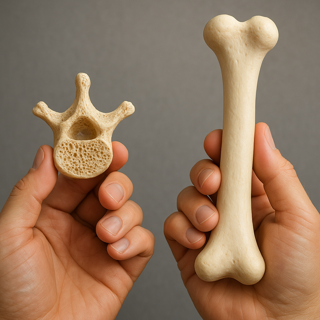
Influence on Bone Microarchitecture
Regular mechanical loading enhances trabecular thickness, connectivity, and alignment along principal stress trajectories. Cortical bone adapts through periosteal expansion and endosteal remodeling, improving cross-sectional geometry. These microarchitectural modifications reduce fracture risk more effectively than increases in mineral density alone.
Adaptation Across the Lifespan
Age and sex significantly modulate the skeletal response to loading. During adolescence, rapid accrual of bone mass and favorable geometry set the foundation for lifelong strength. In adulthood, targeted exercise maintains bone integrity, while in older adults, mechanical stimuli remain effective but require tailored intensity to minimize injury risk. Sex hormones, especially estrogen, modulate mechanosensitivity, explaining differences in adaptation between males and females.
Systemic and Molecular Factors Modulating Adaptation
Bone adaptation to mechanical stress is influenced by a constellation of systemic and molecular factors. Hormonal levels, nutritional status, genetic predisposition, and comorbidities shape the cell’s capacity to sense and respond to mechanical cues.
Hormonal Regulation
Sex steroids, particularly estrogen and testosterone, play pivotal roles in bone homeostasis. Estrogen enhances the proliferation of osteoblasts and inhibits osteoclast formation, promoting an anabolic response to loading. Deficiencies, as seen in menopause or hypogonadism, compromise mechanotransduction efficiency and accelerate bone loss.
Nutrition and Mineral Homeostasis
Optimal bone adaptation requires sufficient calcium and phosphate supply, vitamin D activation, and protein intake. Dietary deficiencies impair mineralization and reduce cellular responsiveness to mechanical stimuli, while excess sodium or caffeine can increase urinary calcium excretion, undermining skeletal adaptation.
Genetic and Epigenetic Influences
Individual variability in genes regulating Wnt signaling, sclerostin expression, and collagen synthesis affects bone’s adaptive potential. Epigenetic modifications, including DNA methylation and histone acetylation, adjust gene transcription in response to mechanical loading history, influencing long-term skeletal health.
Clinical Implications and Therapeutic Strategies
Insights into the bone’s adaptive mechanisms have direct applications in preventing and managing musculoskeletal disorders. Effective interventions integrate biomechanical principles with patient-specific factors such as age, comorbidities, and lifestyle.
Exercise Prescription
- High-impact activities: Plyometric exercises, jumping, and hopping generate ground reaction forces that stimulate bone accrual, particularly beneficial in youth and middle-aged adults.
- Resistance training: Progressive loading through weight machines, free weights, or bodyweight exercises increases cortical thickness and trabecular density at targeted skeletal sites.
- Vibration therapy: Low-magnitude, high-frequency mechanical signals can complement traditional training, especially in populations with mobility limitations.
- Balance and functional exercises: Reducing fall risk through proprioceptive training indirectly protects bone by preventing trauma.
Pharmacological Support
Combining exercise with pharmacotherapy offers synergistic benefits. In osteoporosis management, antiresorptive agents (bisphosphonates, denosumab) and anabolic therapies (teriparatide, romosozumab) enhance the skeletal response to mechanical loading. Coordinated regimens tailored to patient risk profiles maximize bone mass and improve microarchitecture.
Immobilization and Microgravity
Extended bed rest, casting, or microgravity exposure in astronauts leads to rapid bone loss due to lack of mechanical stimuli. Countermeasures include resistive exercise devices, artificial gravity protocols, and pharmacological agents to mitigate disuse osteoporosis. Understanding disuse-induced signaling pathways may reveal novel targets for therapy.
Challenges and Future Directions in Bone Research
The complexity of bone adaptation demands innovative methodologies and interdisciplinary collaboration. Advances in imaging, computational modeling, and biomaterials are paving the way for personalized approaches to skeletal health.
Emerging Technologies
- High-resolution peripheral quantitative CT: Offers three-dimensional assessment of trabecular networks and cortical porosity with micrometer precision.
- Finite element analysis: Integrates imaging data to predict stress distributions and guide individualized rehabilitation protocols.
- 3D bioprinting: Enables fabrication of patient-specific bone scaffolds seeded with osteogenic cells to repair critical defects.
Molecular Interventions
Gene-editing tools such as CRISPR/Cas9 and targeted delivery of RNA molecules hold promise for modulating expression of key mechanosensitive genes. Therapeutics aimed at sclerostin inhibition or enhancement of Wnt signaling may amplify the bone-forming response to mechanical loading.
Translational and Clinical Research
Future investigations will address inter-individual variability in mechanosensing and refine biomarkers that predict adaptive potential. Longitudinal studies combining mechanical assessments, molecular profiling, and functional outcomes are essential for developing personalized exercise and treatment guidelines that optimize skeletal resilience.