Pediatric bone growth and development represent a dynamic interplay of cellular activity, mechanical forces, and systemic influences. Understanding the stages and mechanisms that govern the transformation from a fragile newborn skeleton to a robust adolescent framework is crucial for clinicians, researchers, and caregivers alike. This article delves into the intricate processes that underlie **growth plates**, the role of **osteoblasts**, and the impact of nutrition, genetics, and endocrine factors on skeletal maturation.
Basics of Pediatric Bone Growth
Embryonic Origins and Mesenchymal Differentiation
During the embryonic phase, undifferentiated mesenchymal cells condense and give rise to the structural blueprint of future bones. This condensation process sets the stage for membranous and endochondral ossification routes. In membranous ossification, flat bones such as those of the skull form directly from mesenchyme. Endochondral ossification, which accounts for most long bone formation, involves a crucial cartilage intermediate.
Endochondral Ossification and Cartilage Templates
The cartilage model develops with chondrocytes proliferating at the center, eventually forming a primary ossification focus. Vascular invasion replaces cartilage with mineralized matrix, while the **periosteum** supports collar formation. Secondary ossification centers appear in epiphyses postnatally. The persistence of cartilage at the epiphyseal region allows continued lengthening during childhood and adolescence.
Role of Osteoblasts and Osteoclasts
Bone remodeling is a balance between **osteoblasts**, the builders, and osteoclasts, the resorbers. Osteoblasts secrete osteoid, rich in type I collagen, which later undergoes **mineralization**. Osteoclasts derive from hematopoietic lineages and create resorption pits on old bone surfaces. Coordinated activity ensures structural adaptation to mechanical loads and repair of microdamage.
- Osteoid production and subsequent calcification
- Matrix vesicles initiating hydroxyapatite deposition
- Interplay of RANK/RANKL/OPG signaling in cell regulation
Factors Influencing Skeletal Development
Genetic and Molecular Regulators
Genetic mutations affecting signaling molecules such as fibroblast growth factors and Indian hedgehog can lead to chondrodysplasias and other skeletal dysplasias. Genes controlling collagen synthesis, like COL1A1 and COL1A2, influence **mineralization** and bone strength. Advances in genomics have elucidated pathways that may become therapeutic targets for congenital bone disorders.
Endocrine Influences and Hormonal Modulation
The **endocrine** system orchestrates growth through a symphony of hormones: growth hormone (GH), insulin-like growth factor 1 (IGF-1), thyroid hormones, and sex steroids. GH stimulates chondrocyte proliferation at the growth plates, while IGF-1 enhances matrix production. Thyroid deficiency can retard ossification, and the surge of estrogen at puberty accelerates epiphyseal closure.
- GH/IGF-1 axis in cartilage expansion
- Thyroid hormones modulating chondrocyte maturation
- Sex steroids driving final height determination
Nutrition and Metabolic Considerations
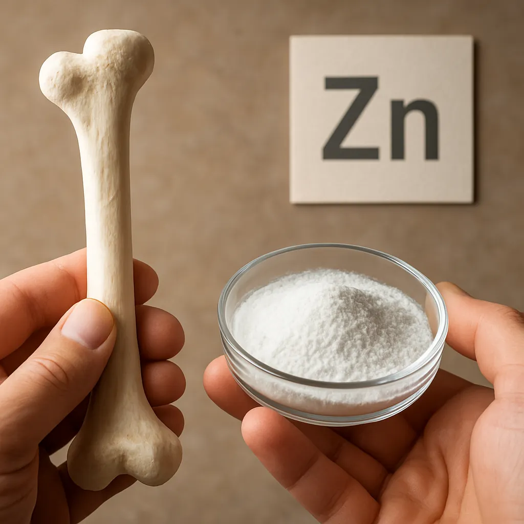
Optimal bone accrual demands an array of nutrients. Calcium and vitamin D are central to **calcification** and mineral homeostasis. Protein supports collagen matrix synthesis. Trace elements like zinc and copper act as cofactors in enzyme systems related to bone formation. Malnutrition, or deficiencies in any of these, can predispose to rickets and weaken structural integrity.
- Calcium absorption enhanced by active vitamin D metabolites
- Dietary protein facilitating osteoid production
- Vitamin K’s role in osteocalcin carboxylation
Mechanical Forces and Wolff’s Law
Bone adapts to the mechanical environment, a principle formalized as Wolff’s Law. Weight-bearing activities stimulate osteoblastic activity and increase bone mass. In contrast, immobilization or lack of mechanical stress leads to resorption and diminished density. Encouraging safe, age-appropriate physical activity in children supports healthy skeletal architecture.
Clinical Approaches and Interventions
Monitoring Growth Plate Health
Evaluation of **growth plates** through radiographic imaging allows assessment of skeletal age and detection of growth disturbances. Early identification of physeal injuries, which can arrest bone growth or cause angular deformities, is paramount. Treatment may involve guided growth techniques such as hemiepiphysiodesis to correct limb discrepancies over time.
Management of Fractures in Children
Unlike adults, pediatric bones possess a thick periosteum and greater remodeling capacity, enabling nonoperative management for many **fracture** types. Greenstick and torus fractures often heal with simple immobilization. Surgical intervention may be required for displaced epiphyseal injuries, using smooth pins or screws to avoid damage to the growth zone.
- Cast immobilization versus operative fixation criteria
- Physeal-sparing surgical approaches
- Rehabilitation protocols to restore function
Pharmacological and Nutritional Therapies
In metabolic bone diseases like osteogenesis imperfecta or juvenile osteoporosis, pharmacological agents such as bisphosphonates reduce bone turnover and increase density. Emerging biologics targeting sclerostin or RANKL show promise. Nutritional supplementation with vitamin D analogues and calcium remains a cornerstone of therapy for rickets and osteopenia.
Innovations in Tissue Engineering and Regenerative Medicine
Advances in stem cell biology and biomaterials open avenues for repairing large bone defects and nonunions. Mesenchymal stem cells seeded onto biodegradable scaffolds can differentiate into osteogenic lineages, promoting **remodeling** and defect bridging. Growth factor–laden hydrogels offer controlled release of osteoinductive signals, enhancing local healing.
Preventive Strategies and Public Health Implications
Public health initiatives targeting childhood nutrition, vitamin D supplementation, and physical activity are vital to reduce the burden of pediatric bone disorders. School-based screening programs for scoliosis and bone density assessment can identify at-risk individuals early. Education of parents and communities about skeletal health fosters environments where children can achieve their genetic potential for strength and growth.