Bone tissue represents a dynamic framework shaped by a delicate interplay between mechanical forces and the endocrine system. Throughout life, bone undergoes constant remodeling, a process essential for maintaining structural integrity and mineral balance. This article examines the intricate network of hormones that regulate bone formation and resorption, explores common metabolic disorders affecting the skeletal system, and highlights emerging therapeutic approaches aimed at preserving and enhancing bone density.
Hormonal Regulation of Bone Remodeling
Bone remodeling is orchestrated by two specialized cell types: osteoblasts, responsible for new bone synthesis, and osteoclasts, which resorb aged or damaged matrix. This continuous cycle ensures adaptability to changing mechanical demands and the preservation of mineral homeostasis. Key endocrine signals modulate each phase of the remodeling sequence, fine-tuning the balance between formation and degradation.
Osteoblast–Osteoclast Interplay
Osteoblast precursors differentiate under the influence of growth hormone and locally produced growth factors such as insulin-like growth factor I (IGF-I). Mature osteoblasts secrete type I collagen and non-collagenous proteins, establishing a mineralized matrix. Simultaneously, osteoblasts express receptor activator of nuclear factor κB ligand (RANKL), which binds to RANK on osteoclast precursors, triggering their maturation into active bone-resorbing cells. Osteoprotegerin (OPG), another osteoblast-derived glycoprotein, acts as a decoy receptor for RANKL, inhibiting osteoclastogenesis and limiting excessive bone resorption.
Endocrine Modulators of Remodeling
- Parathyroid hormone (PTH) enhances osteoclast activity indirectly by upregulating RANKL expression in osteoblastic cells, promoting calcium homeostasis.
- Calcitonin, secreted by thyroid C cells, opposes PTH by inhibiting osteoclast-mediated resorption and increasing renal calcium excretion.
- Sex steroids, notably estrogen and testosterone, suppress osteoclast lifespan and enhance osteoblast function; estrogen deficiency is closely linked to accelerated bone loss.
- Thyroid hormones in excess accelerate both formation and resorption, but net resorptive activity predominates, leading to decreased bone density.
Key Hormones in Bone Metabolism
The skeleton functions as both a structural scaffold and a reservoir for minerals. Its metabolic regulation depends on multiple endocrine axes that adjust bone turnover rates to systemic and environmental demands.
Parathyroid Hormone and Calcium Homeostasis
PTH is secreted in response to hypocalcemia and binds to receptors on osteoblasts and renal tubular cells. In the skeleton, intermittent PTH pulses exert an anabolic effect, stimulating osteoblast proliferation and activity. However, chronic elevation results in pronounced resorption and net bone loss. In the kidneys, PTH increases calcium reabsorption and promotes conversion of 25-hydroxyvitamin D to its active form.
Vitamin D and Mineralization
Vitamin D, synthesized in the skin or obtained from dietary sources, undergoes hepatic and renal hydroxylation to form 1,25-dihydroxyvitamin D (calcitriol). Calcitriol enhances intestinal absorption of calcium and phosphate, essential for hydroxyapatite crystal formation. Adequate vitamin D levels are critical for preventing osteomalacia and facilitating efficient mineralization of the collagen matrix laid down by osteoblasts.
Sex Steroids and Bone Preservation
Estrogen inhibits osteoclastogenesis by upregulating OPG and downregulating RANKL expression. It also promotes osteoblast survival and collagen synthesis. Testosterone supports periosteal bone formation and indirectly contributes to bone mass by its aromatization to estrogen in peripheral tissues. Decline in sex steroid production with aging or surgical removal of gonads significantly increases the risk of osteoporosis and fragility fractures.
Disorders and Clinical Implications
Disruption of endocrine control over bone remodeling can lead to a spectrum of skeletal diseases. Understanding the pathophysiology enables targeted diagnostic and therapeutic strategies.
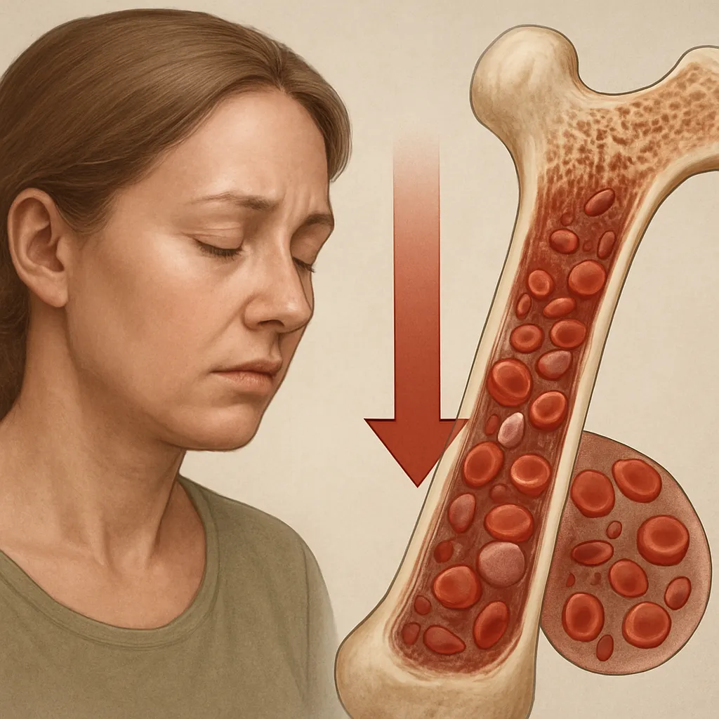
Osteoporosis and Fracture Risk
Osteoporosis is characterized by reduced bone mass and microarchitectural deterioration, resulting in enhanced fracture risk. Primary osteoporosis typically arises from age-related estrogen deficiency in women and relative androgen decline in men. Secondary osteoporosis may result from hyperthyroidism, hyperparathyroidism, glucocorticoid therapy, or malabsorption syndromes.
Hyperparathyroidism and Bone Degradation
Primary hyperparathyroidism involves autonomous overproduction of parathyroid hormone, often due to parathyroid adenomas. Excess PTH causes subperiosteal bone resorption, cortical thinning, and cystic bone lesions. Patients present with hypercalcemia, nephrolithiasis, and skeletal fragility. Surgical removal of the affected gland(s) typically restores bone mass over time.
Rare Endocrine Bone Syndromes
- Hypoparathyroidism: inadequate PTH leads to hypocalcemia, neuromuscular irritability, and impaired bone formation, manifesting as increased density but poor quality.
- Paget’s disease of bone: focal overactivity of osteoclasts and disorganized osteoblastic repair resulting in enlarged, brittle bones.
- Cushing’s syndrome: chronic glucocorticoid excess diminishes osteoblastogenesis and accelerates osteocyte and osteoblast apoptosis, promoting osteoporosis.
Therapeutic Approaches and Future Directions
Modern interventions aim to restore the equilibrium between bone formation and resorption while minimizing adverse effects. Pharmacologic agents, lifestyle modifications, and investigational biologics all contribute to comprehensive bone care.
Anti-Resorptive and Anabolic Agents
- Bisphosphonates: inhibit osteoclast-mediated resorption by inducing apoptosis in active osteoclasts, reducing fracture incidence.
- Denosumab: a monoclonal antibody against RANKL that prevents osteoclast differentiation and activity.
- PTH analogs (teriparatide): intermittent administration stimulates osteoblastic bone formation and increases bone mass.
- Selective estrogen receptor modulators (SERMs): mimic estrogen’s protective effects on bone without stimulating breast or uterine tissue.
Lifestyle and Nutritional Strategies
Adequate dietary intake of calcium and vitamin D, weight-bearing exercise, smoking cessation, and moderation of alcohol consumption are foundational measures. Fall prevention programs and balance training further mitigate fracture risk in the elderly.
Emerging Research and Regenerative Medicine
Novel therapies under investigation include sclerostin inhibitors, which lift the brake on bone formation imposed by the Wnt signaling antagonist sclerostin, and cell-based approaches utilizing mesenchymal stem cells to replenish osteoblast populations. Advances in molecular genetics are uncovering polymorphisms in genes encoding calcitonin receptors and vitamin D-binding proteins, paving the way for personalized bone health management.
By integrating insights from endocrinology, molecular biology, and biomechanics, clinicians and researchers continue to refine strategies that preserve skeletal strength and quality of life across the lifespan. Ongoing collaboration between basic science and clinical disciplines promises to unlock innovative modalities for optimizing bone health under diverse physiological and pathological conditions.