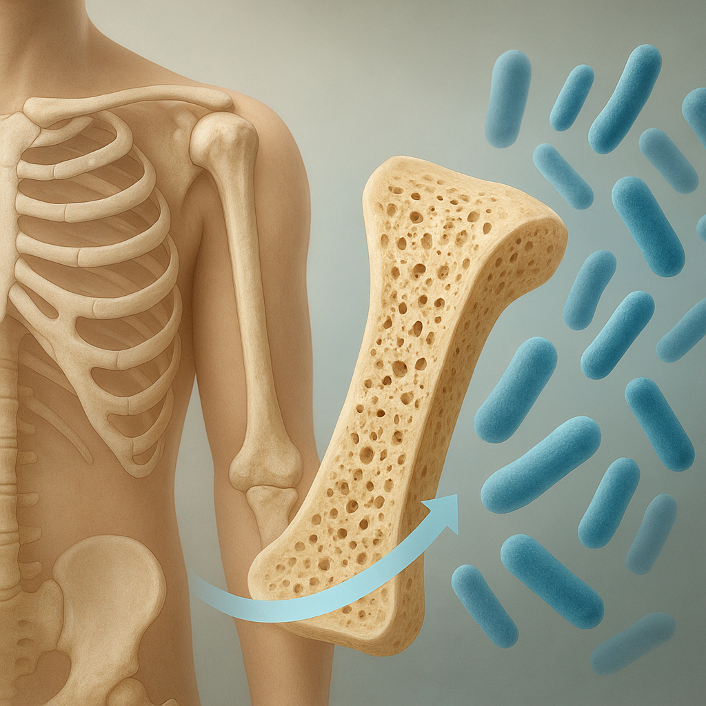
Bone scaffold engineering has emerged as a pivotal field within orthopedics and regenerative medicine, aiming to restore dysfunctional skeletal tissues through innovative designs and materials. By integrating advances in biomaterials science, cutting-edge fabrication methods, and cellular biology, researchers strive to create constructs that not only support mechanical loads but also guide cellular processes like osteogenesis and tissue remodeling. This article delves into the latest developments across material selection, manufacturing approaches, and biological integration strategies, highlighting the challenges and opportunities that shape the future of bone repair technologies.
Scaffold Materials and Design
The choice of base materials is fundamental to scaffold performance. Natural polymers, such as collagen and chitosan, offer inherent biocompatibility and biomimetic cues but may lack mechanical strength. Synthetic polymers like poly(lactic-co-glycolic acid) (PLGA) and polycaprolactone (PCL) deliver tunable degradation rates and robust load-bearing capacity. Combining these with inorganic fillers, such as hydroxyapatite or bioactive glass, yields composite systems that bridge the gap between mechanical resilience and biological function.
One promising route involves creating nanocomposites by embedding nanoscale particles into polymer matrices. These particles enhance surface roughness and present osteoinductive signals at the cellular interface. Additionally, tailoring scaffold porosity and pore interconnectivity is crucial, as it dictates nutrient diffusion, cell migration, and vascular ingrowth. Porous architectures ranging from 50 to 500 micrometers are often preferred to optimize bone tissue infiltration and new vessel formation.
Advanced computational modeling aids in predicting mechanical performance under physiological loads. Finite element analysis (FEA) guides design iterations, ensuring that stress distributions within the scaffold mimic those of native bone. Topology optimization can generate structures with precise control over local stiffness, enabling regions of high strength and zones that encourage controlled deformation to stimulate mechanotransduction pathways.
Additionally, surface functionalization techniques—such as peptide grafting or growth factor immobilization—provide targeted biochemical signals. Incorporating motifs like RGD (arginine-glycine-aspartate) enhances cell adhesion, while tethered bone morphogenetic proteins (BMPs) accelerate differentiation of progenitor cells. Such strategies integrate the dual demands of mechanical support and bioactivity for optimal healing outcomes.
Fabrication Techniques
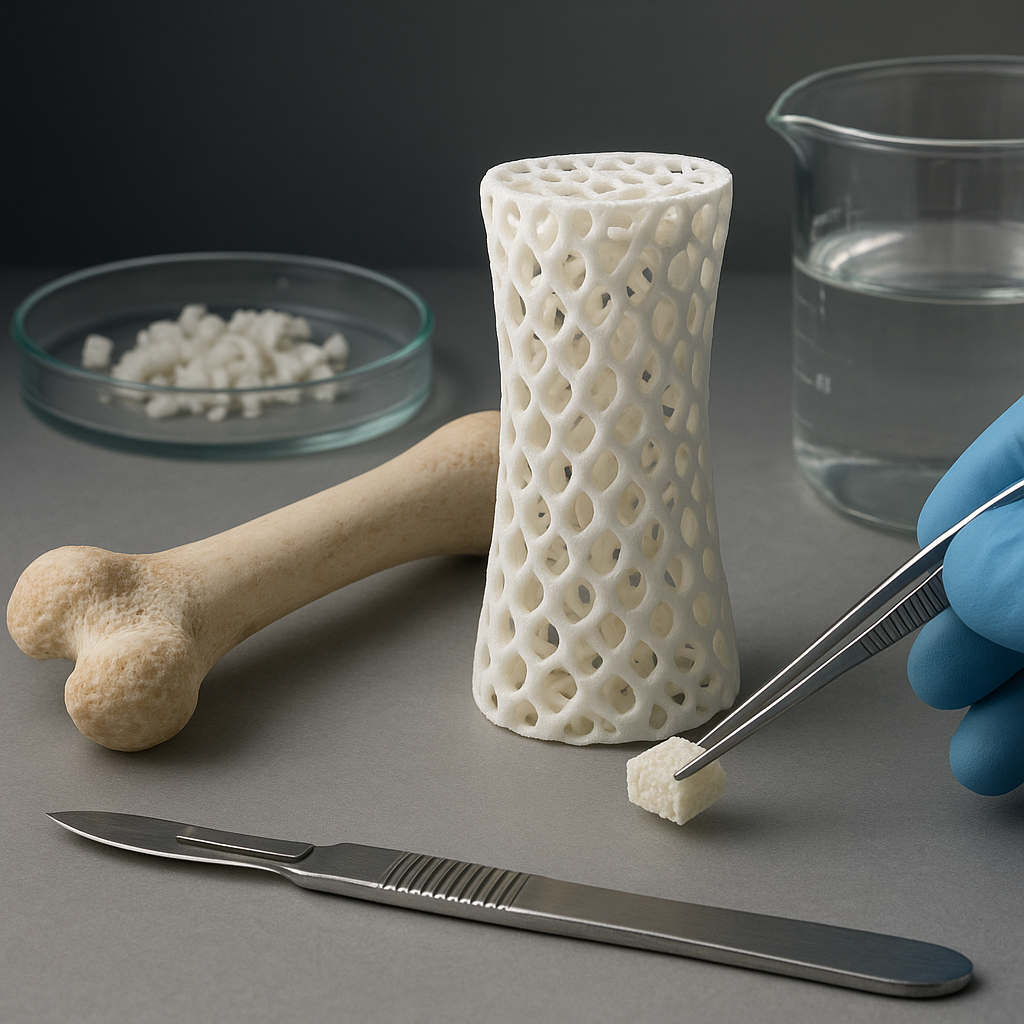
The advent of additive manufacturing has revolutionized scaffold fabrication by enabling patient-specific geometries and reproducible architectures. 3D printing methods, including fused deposition modeling (FDM), stereolithography (SLA), and selective laser sintering (SLS), offer distinct advantages for multimaterial constructs. FDM can process thermoplastic polymers with embedded ceramic powders, while SLA achieves high-resolution features using photosensitive resins.
Robocasting and direct ink writing (DIW) techniques extrude viscous bioinks to build layered structures at ambient conditions, preserving the viability of encapsulated cells or growth factors. These extrusion-based methods facilitate the incorporation of hydrogels, enabling the creation of hybrid scaffolds with soft and rigid domains. Layer-by-layer deposition ensures precise control over pore size and strut thickness, essential for balancing mechanical integrity and mass transport.
Electrospinning remains a versatile approach for generating fibrous scaffolds that mimic the extracellular matrix. By controlling solution viscosity, electric field strength, and collector geometry, researchers can produce aligned or random fiber mats with nanoscale diameters. These mats can be stacked or integrated with 3D-printed frames to combine nanoscale topography with macroscopic support.
Emergent techniques like 4D printing introduce responsiveness to external stimuli. By using shape-memory polymers or hydrogel composites, scaffolds can change shape upon temperature shifts or hydration, enabling minimally invasive implantation and subsequent expansion within defect sites. Such dynamic constructs promise improved conformity to irregular bone cavities and enhanced mechanical anchorage.
Biological Integration and In Vivo Performance
Successful bone regeneration hinges on the scaffold’s ability to recruit cells, support differentiation, and foster vascularization. Pre-seeding constructs with mesenchymal stem cells (MSCs) provides a living component that secretes osteogenic factors and remodels the matrix. Co-culturing MSCs with endothelial cells further stimulates vascularization, ensuring adequate blood supply to the developing tissue.
Growth factor delivery remains an area of intense research. Controlled-release systems embed BMP-2, vascular endothelial growth factor (VEGF), or platelet-derived growth factor (PDGF) within micro- or nanoparticles dispersed throughout the scaffold. This spatiotemporal presentation of signaling molecules guides cell behavior, mitigating the risks of burst release and off-target effects.
Animal studies have demonstrated that implants featuring graded mechanical properties—stiffer at load-bearing surfaces and more compliant at interfaces—promote gradual load transfer to regenerating bone. Monitoring in vivo degradation profiles is essential to ensure scaffold resorption aligns with new tissue formation. Polymers or composites with tailored biodegradability enable safe byproduct clearance without inflammatory responses.
Biomechanical testing post-implantation assesses parameters like compressive strength and elastic modulus recovery. Histological analyses reveal the extent of mineral deposition, collagen matrix organization, and vessel infiltration. Collectively, these data inform iterative improvements, moving from small rodents to large animal models before clinical translation.
Emerging Trends and Future Directions
Integration of digital healthcare tools is shaping personalized scaffold design. Imaging modalities such as CT and MRI provide high-resolution defect mapping, feeding directly into computer-aided design (CAD) workflows. Patient-specific implants optimize fit and distribution of mechanical loads, reducing the risk of stress shielding and implant failure.
Bioprinting, which combines living cells with hydrogel matrices, is advancing toward fully cellularized bone grafts. Recent breakthroughs in microfluidic printheads enable the deposition of multiple cell types and growth factors in precise spatial patterns, mimicking native bone marrow niches. This approach holds promise for repairing critical-sized defects and non-union fractures.
The convergence of gene editing and scaffold engineering offers new avenues for enhancing regenerative capacity. Scaffolds can deliver plasmids or viral vectors encoding osteogenic transcription factors directly to stem cell populations in situ, boosting their differentiation potential. Such gene-activated matrices may reduce the need for exogenous growth factors and simplify regulatory pathways.
Finally, mechanobiology-driven designs seek to harness every day physical activities. Smart scaffolds equipped with piezoelectric components generate electrical cues in response to mechanical loading, further stimulating cellular functions. By coupling electrical and biochemical signals, these next-generation constructs aim to replicate the complex milieu of the bone microenvironment, ushering in a new era of functional bone tissue engineering.